
Tomorrow is my grandmother’s 103rd birthday. So this article is for her in more ways than one.
Upon further contemplation, I decided to write this article in two parts, covering two distinct yet highly significant papers.
Before I republish my second Language article, while I steadily work on the third (the new article, and now the fourth and fifth…lol), I decided to write this article, which, from my perspective, is a better fit to publish before the one focusing on microstructural and microvascular/capillary damage, because it gives a large basis for multiple observations from distinct points regarding SARS-CoV-2.
Towards that goal, I will write a brief explanation of the main subject and also my own observations over the years. The main subject will be one specific Interferon. So, what are interferons ?
Interferons are a class of cytokines, proteins that function as chemical messengers, enabling cells to signal one another and coordinate responses. They are antiviral by nature, though also released in response to bacterial detection. When a cell encounters a pathogen or antiviral signal, it releases interferon, thereby signaling nearby cells to prepare their defenses, and actively disrupting replication of pathogens inside the cells. Interferons (IFN) are powerful initiators and orchestrators of immune responses, and their presence initiates the transcription of hundreds of genes. They are divided into 3 classes.
Type 1 Interferon consists of IFN-α, IFN-β, IFN-ε, IFN-κ, and IFN-ω, with the most common being Alpha and Beta.
Type 3 Interferon consists of a family sharing the same name λ, meaning Lambda, with λ1, λ2, λ3, λ4. Its main role is protecting epithelial cells in the lungs, gut, and skin.
And lastly, we have our focus - Type 2 Interferon. IFN-γ (Gamma), being the single Interferon in this class.
IFN-γ (I will sometimes call it Gamma from here on, for simplicity) is of special interest for a multitude of reasons. Primarily released by T-Cells and Natural Killer cells, it works at very low concentrations, so it is fairly potent. Gamma’s functions include activating macrophages and increasing antigen presentation, it can promote Th1 immunity, shape leukocyte recruitment, influence endothelial activation and permeability, modulate neuroinflammation, modulate systemic immune responses, and more.
Possessing such potency, to avoid a runaway effect of inflammation, the body has a few brakes for it, with the main one being widely recognizable if you are a long-time reader. IDO, the primary enzyme responsible for initiating the infamous Kynurenine Pathway.
One core part of my general hypothesis regarding all forms of damage and sequelae from SARS-CoV-2 and its fragments has been that Gamma and its superfamily of interleukins play a disproportionate and paradoxical role in both damage and long-term consequences of SARS-CoV-2. So much so that I was already using Gamma as a biomarker for Long Covid (specific subtypes) back in 2021. It is the very definition of a double-edged sword, as much as the immune system needs it, a higher concentration or persistent release at low concentration induces damage, and from my research, the effects were always systemic, and responses were not localized.
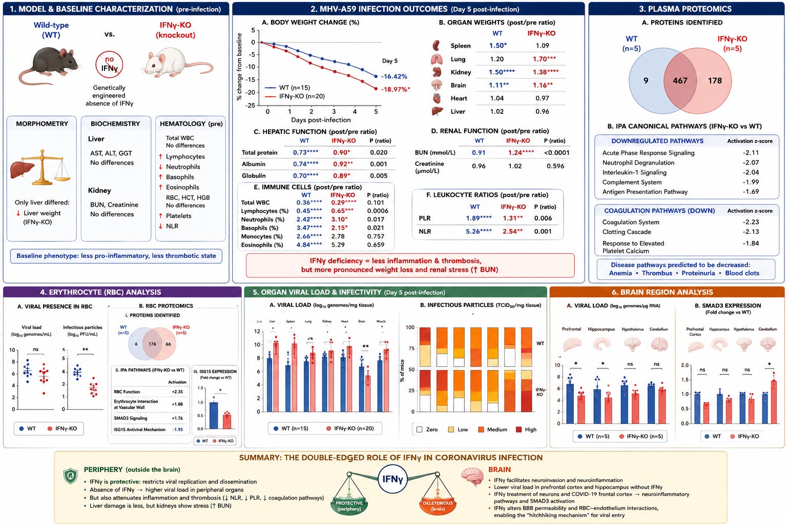
A summary in illustration form from the paper we are about to cover.
Throughout the years, we have had a number of papers on the subject, with a focus on the dysregulation of Gamma in Covid patients, but Gamma remains understudied regarding its role in the pathogenesis.
The authors start by comparing two models. “Normal” mice (Wild-type, WT) and IFNγ-KO (knockout) mice, which are genetically engineered to have no IFNγ whatsoever. This allows them to isolate the consequences of Gamma absence during a live coronavirus infection. Before infection, the two types of mice were broadly similar. Organ-wise, only the liver differed, weighing slightly less in the KO mice, liver markers such as AST, ALT, and GGT, and renal function markers were all comparable between the two.
The distinction starts in the immune system, total White Blood Cells didn’t differ, but the Gamma-KO mice had increased lymphocytes, basophils, and eosinophils, while possessing fewer neutrophils. Hematological (blood) profiling showed no specific changes, with comparable Red Blood Cell count, hematocrit, or hemoglobin. However, IFNγ-KO mice had increased platelet numbers and a decreased neutrophils-to-lymphocytes ratio (NLR), this is a common marker for systemic inflammation. This baseline profile is important as it establishes that IFNy deficiency creates a less pro-inflammatory, less thrombotic state.
With a delineated baseline, the authors proceed to test their hypothesis by using Murine Hepatitis Virus A59(MHV-A59), a betacoronavirus widely used to model coronavirus disease, especially SARS-CoV-2.
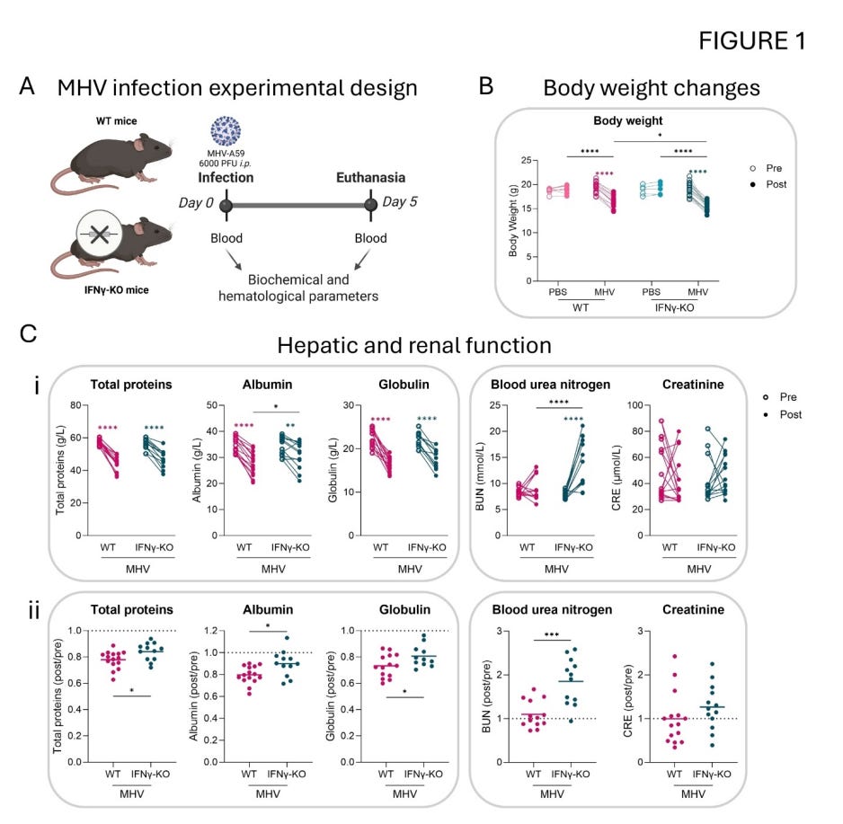
Body weight change is a widely used marker for acute illness, both in research and clinical settings, and here Gamma-KO mice experienced a more pronounced weight loss compared to WT-infected controls. Organ-wise, MHV infection increased spleen weight in the WT group (showing immune activation) and lung weight in the Gamma-KO group (evidence of distinct inflammatory patterns), kidney and brain presented higher weights in both. Heart and liver weights and hematological markers didn’t show differences.
Upon assessing hepatic function, they found MHV infection significantly reduced total protein, albumin, and globulin in both genotypes, albeit these reductions were significantly less pronounced in the Gamma-KO group, meaning reduced hepatic (liver) damage. No significant differences in liver enzymes in both. In renal function, the IFN-Gamma-deficient group experienced a significant increase in BUN (a marker for kidney function and protein catabolism), indicating that while the liver is spared, the kidneys enter a stressed compensatory state.
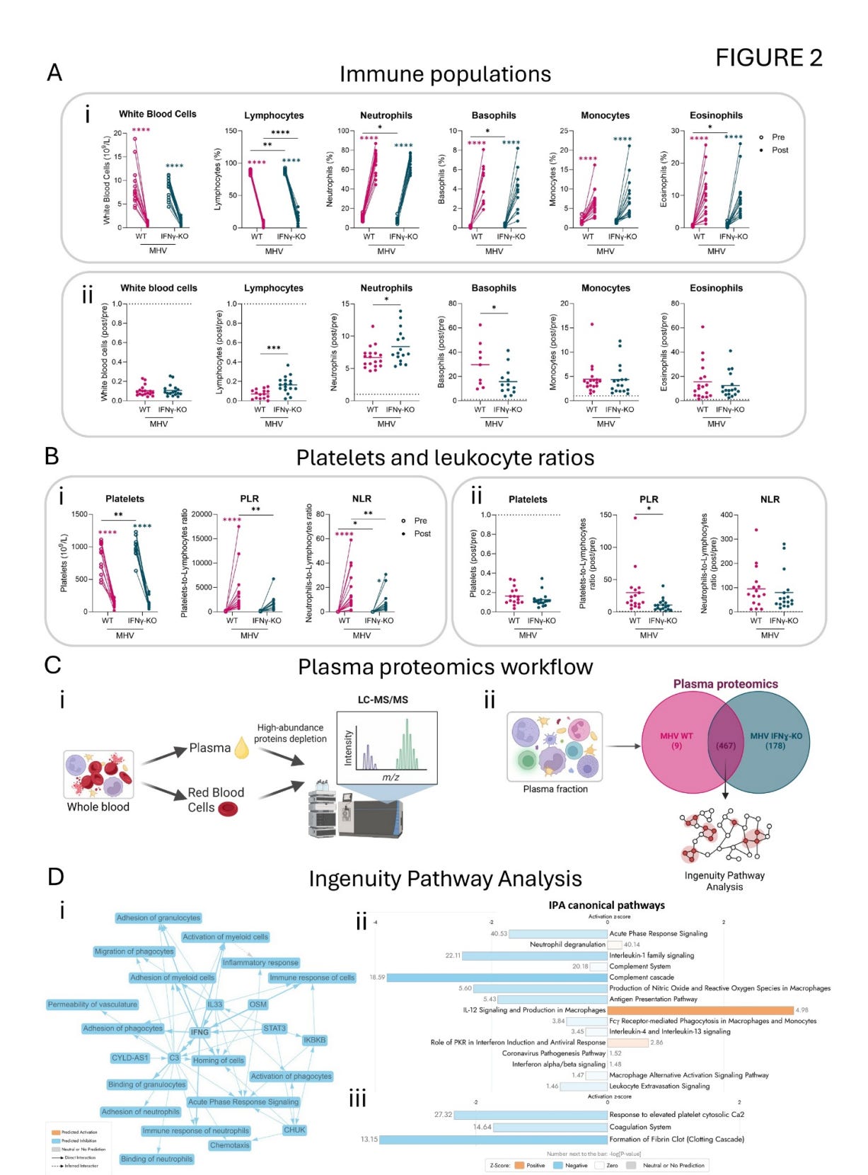
When analyzing immune parameters, the infection caused a decrease in total White Blood Cell numbers and in lymphocyte percentage in both groups, while an increase in neutrophils, basophils, monocytes, and eosinophils was observed in both after infection. Comparing post- and pre-infection ratios between genotypes, Gamma-KO mice presented a smaller reduction in lymphocytes, less augmented basophils, but a higher increase in neutrophils, showing a distinct immune strategy when Gamma is absent.
Platelets and leukocytes ratios (PLR, NLR, respectively) were decreased in Gamma-KO. This is relevant because both PLR and NLR are reliable biomarkers for poor outcomes in viral infections. High NLR reflects increased neutrophil infiltration and reduced lymphocyte-mediated antiviral response, while elevated PLR indicates a prothrombotic state. This paints a picture in which IFNγ drives both inflammatory and thrombotic responses during a coronavirus infection.
The next step is to measure the proteins in the blood plasma (plasma proteomics) to analyze the differences between the mice without IFN-γ and WT. 654 proteins were identified in total, with 467 shared between genotypes. 178 proteins were unique to IFNy-KO, and only 9 were unique to WT. Using IPA (Ingenuity Pathway Analysis, extremely good software for predicting and connecting pathways from data like this one) revealed a lot about the lack of IFNγ.
Immune pathways such as Acute Phase Response Signaling, Neutrophil Degranulation, Interleukin-1 signaling, Complement System, and Antigen Presentation Pathway were all downregulated. Coagulation System, Clotting Cascade (such as the formation of fibrin clots), response to elevated platelet calcium were negatively regulated. Disease pathway analysis further predicted decreased infection-related and blood-related pathologies such as anemia, thrombus, and proteinuria in the KO animals.
One dynamic shared by some coronaviruses such as, MHV (one of the previous research from this group) and SARS-CoV-2, is that they change the fate of Red Blood Cells (RBCs). SARS-CoV-2 is especially significant because it infects and affects the precursors of RBCs. Although they found no significant differences in viral loads, they did observe that infectivity was reduced in RBCs of the IFNγ-deficient mice.
This distinct pattern is significant as it suggests that IFNγ does not just regulate viral replication in RBCs, but influences the quality, packaging, and stability of infectious virions associated with these cells, or modulates RBC-viral interactions that preserve infectivity. Given that IFNγ can affect endothelial adhesiveness and vascular permeability, it impacts RBC-endothelium interactions that facilitate viral spread, a dynamic the authors previously termed the “hitchhiking mechanism”.
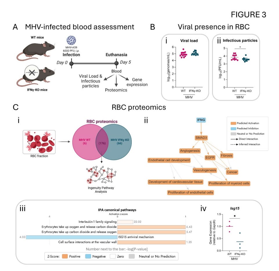
Performing the same proteomic analysis on the Red Blood Cells, they identified 248 proteins, 176 of which were shared between both genotypes. Pathway analysis confirmed Gamma deficiency and found something new and unexpected, increased SMAD3 signaling. SMAD3 is involved in brain injury protection, fibrosis, and angiogenesis, and mediates endothelial permeability. This is a very impactful signal in this paper. The same analysis indicated enhanced RBC function and interactions at the vascular wall in the mice without IFNγ.
Paradoxically, the RBC proteomics also indicated repressed ISG15. Interferon-stimulated gene 15 is canonically considered a Type I IFN-stimulated gene with broad antiviral activity. Its downregulation in IFNγ-KO RBCs, despite the presence of Type I IFN expression elsewhere in the body, suggested that IFNγ is significantly necessary for full ISG15 induction.
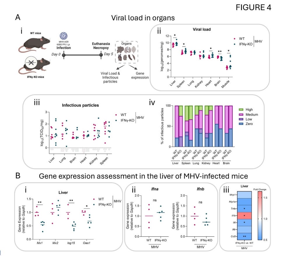
The effects of IFNγ deficiency are rather drastic. It leads to systemic viral dissemination, especially in the liver (MHV’s primary target), spleen, heart, and muscle, yet it significantly spares the brain, which was unexpected. So next, they examined the proteomic signatures associated with neurological processes and found processes related to neurological diseases, neuronal cell death, immune activation, and bleeding were all negatively regulated, meaning the brain was protected in the KO mice.
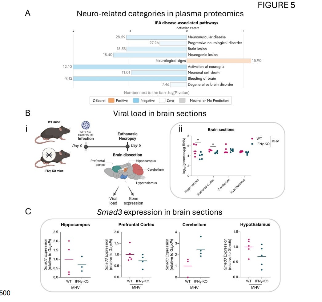
What to be is another insightful part of this research was the dissection of the brains after infection, because it showed something unique. Distinct parts of the brain were protected, the viral load was significantly lower in the prefrontal cortex and hippocampus, but no differences were seen in the hypothalamus or cerebellum in the Gamma-deficient mice.
Since SMAD3 was predicted to be activated in the RBCs, they assessed its expression across different parts of the brain. SMAD3 was reduced in the hippocampus, prefrontal cortex, and hypothalamus of Gamma-deficient mice, but more pronounced in the cerebellum.
The data indicate that IFNγ has a region-specific effect. Worth noting that while SARS-CoV-2 impacts the whole brain via numerous mechanisms, two of the most affected regions are the prefrontal cortex and hippocampus. But does all the data and changes observed here relate to “actual” infection in humans ?
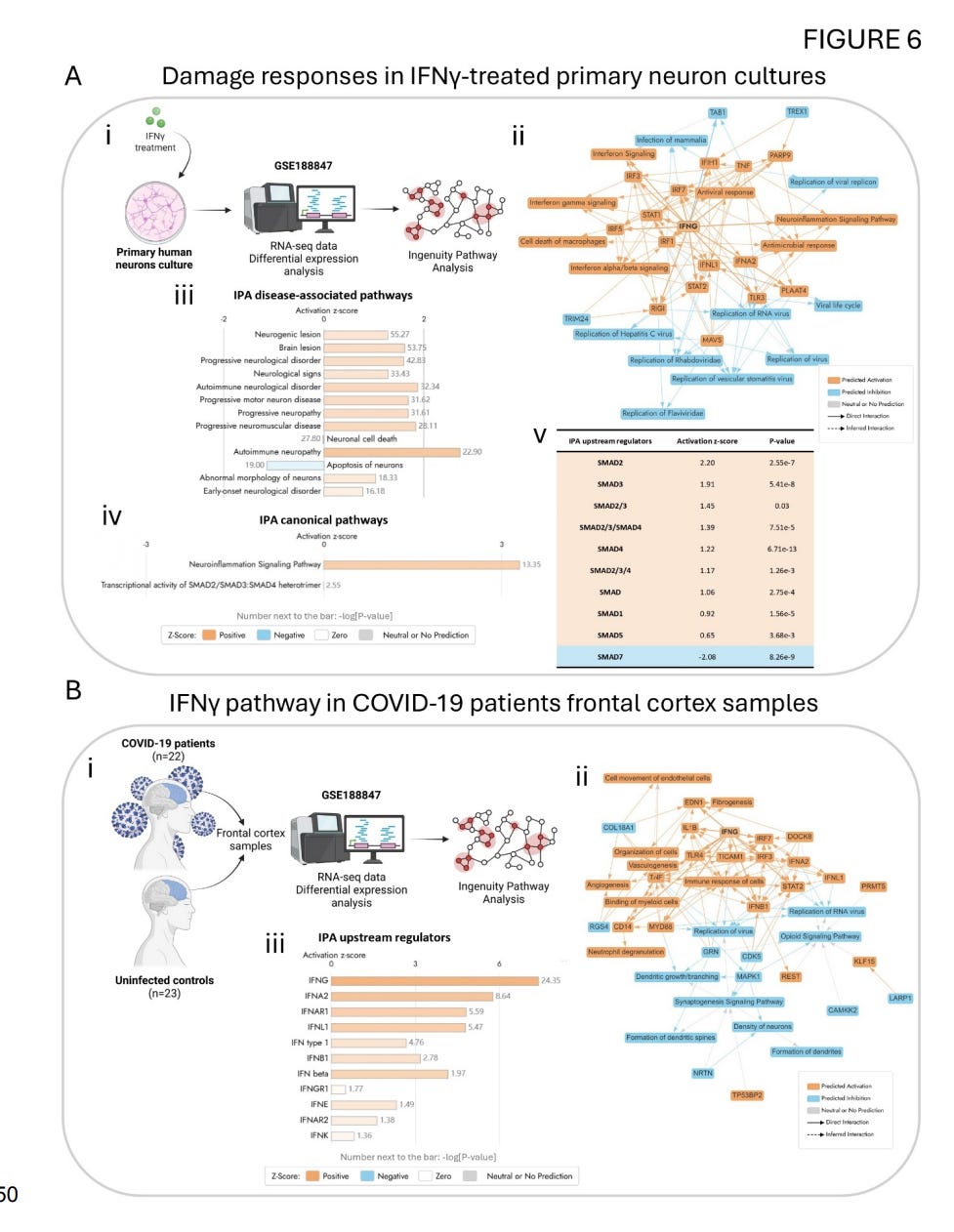
To pursue this assessment, the authors analyzed publicly available datasets from neurons treated with IFNγ and from the frontal cortex of Covid patients. The treatment of neuron cultures with IFNγ increased the expression of genes related to neuroinflammation and antiviral responses.
In neuron culture data, IFNγ treatment increased the expression of genes related to neuroinflammation and antiviral responses. Disease pathway analysis detected dysregulations overlapping with brain lesions, progressive neurological disorders, and neuronal cell death. Among the canonical pathways, Neuroinflammation Signaling and the transcriptional activity of SMAD2/SMAD3:SMAD4 were significantly activated. Upstream regulator analysis further identified SMAD3 as the second most activated member of the SMAD family in responses to IFNγ.
Turning to the patient data, the frontal cortex of Covid patient showed predicted activation of the IFNγ signaling pathway. This was associated with increased fibrogenesis and immune responses, but decreased viral replication and formation of neural networks. Crucially, among all interferons analyzed, those being IFNγ, IFNα, IFNAR1, IFNλ, and others, IFNγ had the highest activation z-score in the frontal cortex, far exceeding both Type I and III interferons.
This research provides contextually significant findings on the double-edged nature of IFNγ. In the periphery (outside the brain), Gamma is protective, it synergizes with Type I interferons to drive maximal ISG expression in many organs. Without it, viral replication goes unchecked. However, in the absence of Gamma, it also remarkably attenuates inflammatory and thrombotic responses systemically.
In the brain, the role is reversed. Gamma can facilitate neuroinvasion by changing Blood-Brain Barrier permeability, altering junctional integrity, and creating entry points for the virus or inflammatory mediators. It enhances RBC-endothelial interactions and alters vascular permeability, facilitating the “hitchhiking” mechanisms allowing infectious particles to access the neurovasculature.
This article is already lengthy, and even though I have aimed to present the information and data in a manner that most people can understand, adding references to my other articles, creating a complex chain of references, would extend the length to an uncomfortable level and add to the complexity. So if you are interest into going into the unknown, use the search tool on my main page, and you will get numerous articles.
Together with a broader set of clinical, immunological, and observational data from a myriad of sources, with the latest addition providing significant insight, we understand IFNγ is central to coronavirus responses. Alone, this interferon has a remarkable footprint on almost every part of the body, it participates in and influences hormonal responses together with other inflammatory proteins, it affects the bone marrow, directly interacts with the HPA axis (Hypothalamic-Pituitary-Adrenal), the main neuroendocrine axis governing systemic stress responses and cortisol release.
In Covid, HPA axis activation can occur even before fever onset, driven in part by IFN-γ alongside IL-6 and TNF-α. Gamma is powerful enough to antagonize the establishment of endotoxin tolerance, the immuneparalytic state seen in severe sepsis, late-stage critical illness, and post-severe Covid, effectively reversing a state of immunosuppression.
This creates a double-hit effect. In an attempt to limit the effects of IFN-γ, the Kynurenine Pathway kicks in, and ironically enough, while this effectively keeps Gamma in check, it contributes to a paradoxical state of chronic, sometimes “sterile” inflammation. IDO1 activation depletes tryptophan from the microenvironment at first, and this inhibits T-Cell proliferation, creating a immunosuppresive state.
Kynurenine metabolites exert diverse, and often paradoxical effects. 3-Hydroxykynurenine generates oxidative stress and contributes to endothelial damage. Quinolinic acid is neurotoxic, but it does so by acting as an NMDA receptor agonist, driving excitotoxicity and neuronal injury through overactivation of glutamate signaling. Kynurenine acid, in contrast, is neuroprotective via NMDA receptor antagonism and anti-inflammatory properties, but in Covid, the pathway shifts towards quinolinic acid production.
Exposure to any amount of IFNγ is able to induce profound, lasting changes in immune responses via epigenetic changes, a process called epigenetic inflammatory memory, where your body remembers the inflammatory response. In monocytes and macrophages, IFNγ primes inflammatory gene loci through histone modifications such as H3K4me3 and H3K27ac, creating a chromatin state that enables faster, stronger responses to subsequent stimuli.
In a disease-dependent manner, IFNγ can drive persistent epigenetic shifts and contribute to damage to the synapses. With contributing factors, IFNγ is able to induce IDO1 expression via an epigenetic mechanism. Solving the epigenetic memory puzzle with regard to viral infection sequelae is essential, as the epigenetic scars left by these processes (histone modifications, altered chromatin landscapes, promoter methylation changes, and metabolic rewriting) outlast the virus. SMAD3-dependent neurovascular modulation described in the primary paper discussed earlier likely represents one downstream node of this broader pathway.
This is an effort towards both understanding what is underlying the long-term consequences of Covid, but also unveiling hidden mechanisms so we can properly heal people. Clinically speaking, without an assortment of biomarkers, in the presence of elevated Gamma, you should first target the gut and brain, followed closely by endothelial health. A variable is the presence or detection of antibodies against Herpesviruses and other latent viruses, in this case, targeting them also becomes essential.
Thank you for your continued support, it is very helpful. Consider becoming one or you can buy me a coffee as a one-time thing.
No posts
Comments
Nothing yet. Say the first thing.
Sign in to join the conversation.