
A woman walks into clinic having already seen several IVF specialists. Two years of primary infertility, and an HSG report reading “bilateral tubal blockage.” By the time patients like this reach a gynaecologic surgeon, the default pathway has usually already been proposed to them: skip the tubes, go straight to IVF.
The numbers explain why. Infertility affects an estimated 10–15% of couples worldwide — by some estimates, as many as 1 in 6. Endometriosis affects roughly 5–10% of women of reproductive age.
On their own, those are two moderately common conditions. But they are not independent of each other: among women undergoing laparoscopy specifically for infertility evaluation, published series put the prevalence of endometriosis dramatically higher — commonly cited in the range of 25–50%, several-fold above the background population rate.
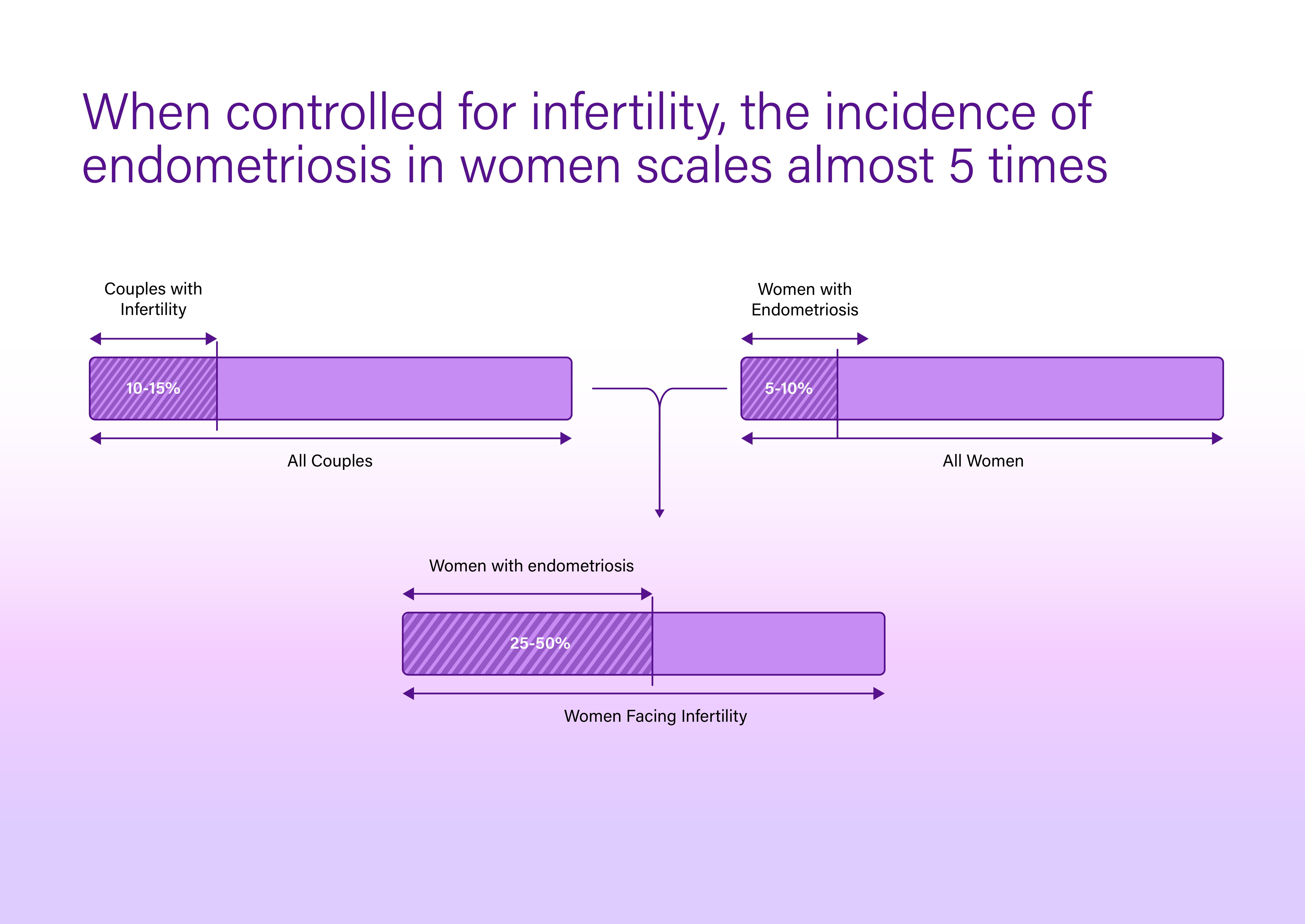
The two conditions travel together often enough that a surgeon who ignores the possibility of coexisting endometriosis in an infertile patient is working with half the picture.
But the more interesting question isn’t whether disease is present. It’s what to do once you find it. This week’s case is exactly that scenario — and the lesson it offers is less about what we did than about what we deliberately chose not to do.
A patient with two years of primary infertility, multiple prior fertility consultations, and an HSG demonstrating bilateral tubal blockage was referred to us. Consultation, history, examination, and diagnostic workup followed, and we proceeded to combined hysteroscopy and laparoscopy.
Hysteroscopic findings were normal. The tubal blockage seen on HSG, however, needed to be addressed directly. Using a guidewire under hydrostatic pressure, both tubes were recanalised, followed by chromopertubation. Dye flowed freely on both sides — the obstruction identified on imaging was resolved at the level accessible hysteroscopically.

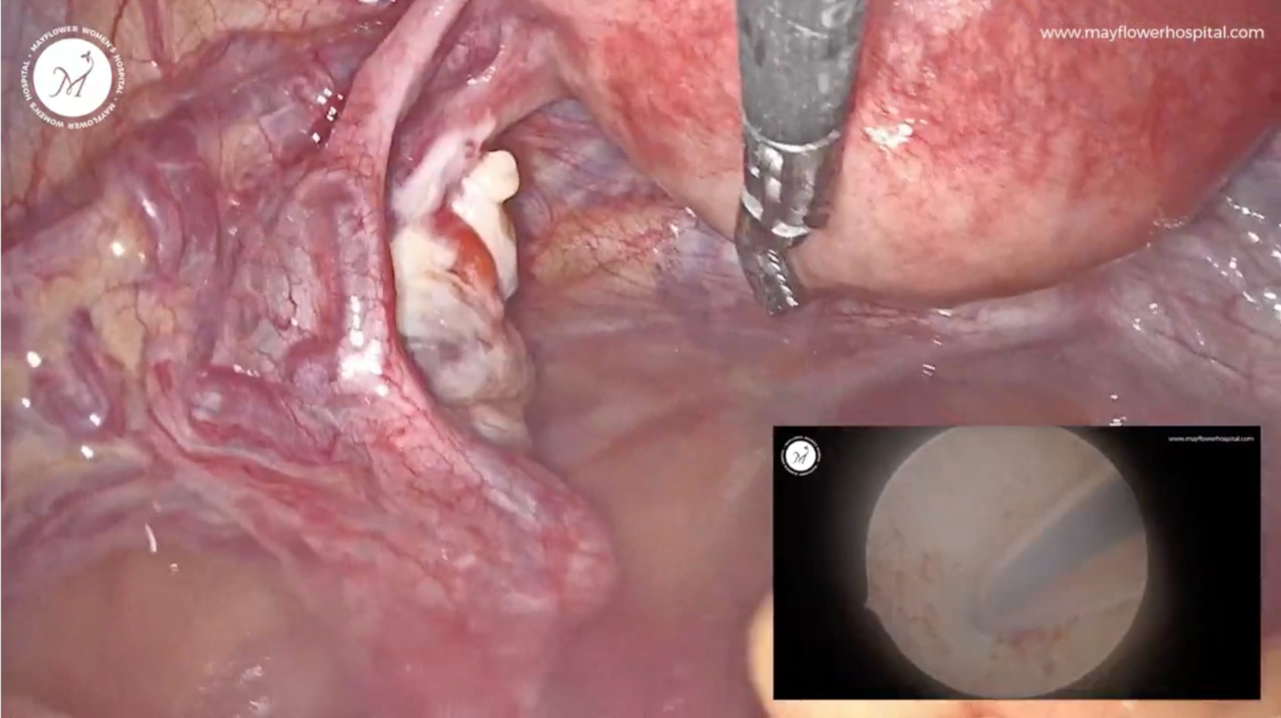
Guidewire cannulation of the tubal ostium under hydrostatic pressure; inset shows the hysteroscopic view.
On entering the abdomen, three sites of disease were apparent: bilateral parametrial involvement over the uterosacral ligaments, a rectovaginal plaque, and a plaque over the bladder serosa.
On both sides, the disease sat at a comfortable distance from the ureter. That single observation changed the operative plan. Rather than defaulting to full bilateral ureterolysis — standard in more extensive deep disease — we kept the ureter in view and protected, and confined dissection to the plaque itself. Each parametrial plaque was excised locally, ureter safeguarded, on both sides in turn.
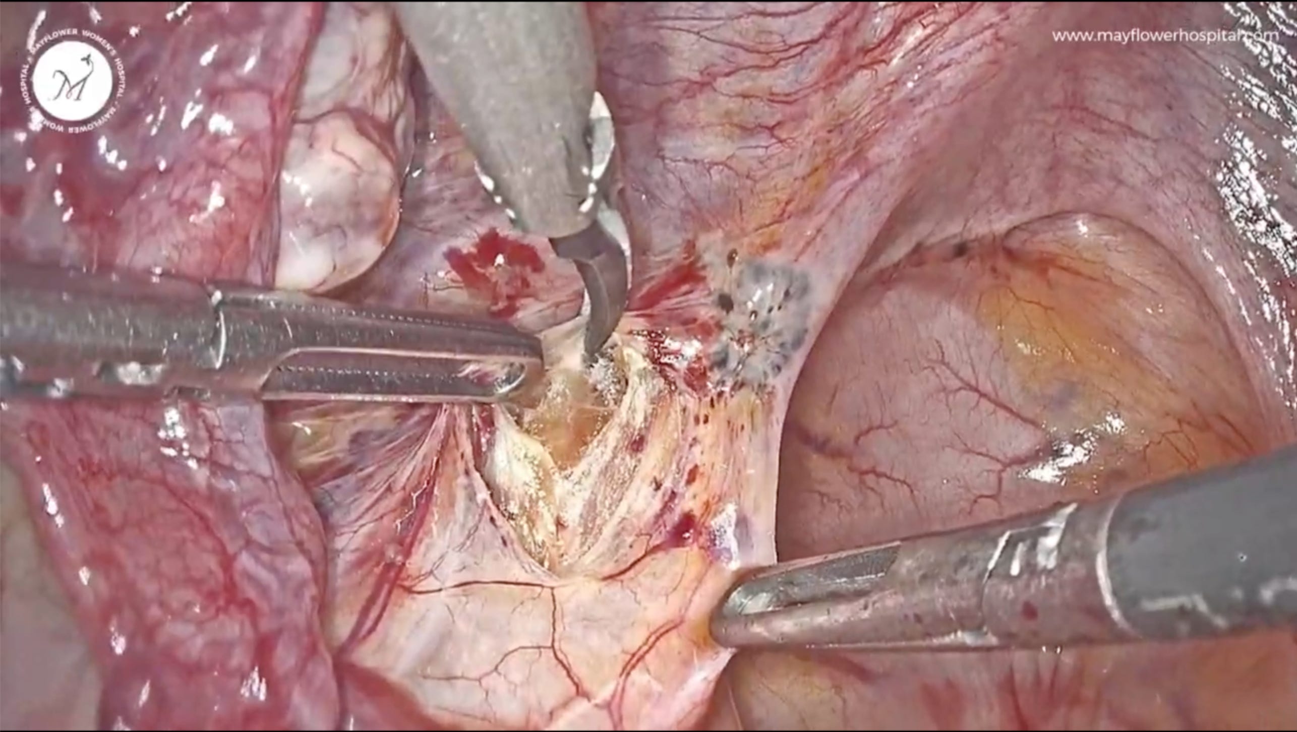
Targeted excision of parametrial disease. Full ureterolysis was deliberately not performed on either side, since the disease did not approach the ureter.*
Attention turned to the rectovaginal plaque. Dissection entered the correct plane of Denonvilliers’ fascia, the rectum was mobilised and safeguarded, and the disease was removed from that plane.
The bladder plaque presented as a nodule between the left round ligament and the bladder wall. It was grasped firmly, the surrounding loose tissue mobilised, and the plaque removed fiber by fiber off the bladder serosa — preserving the integrity of the underlying wall.
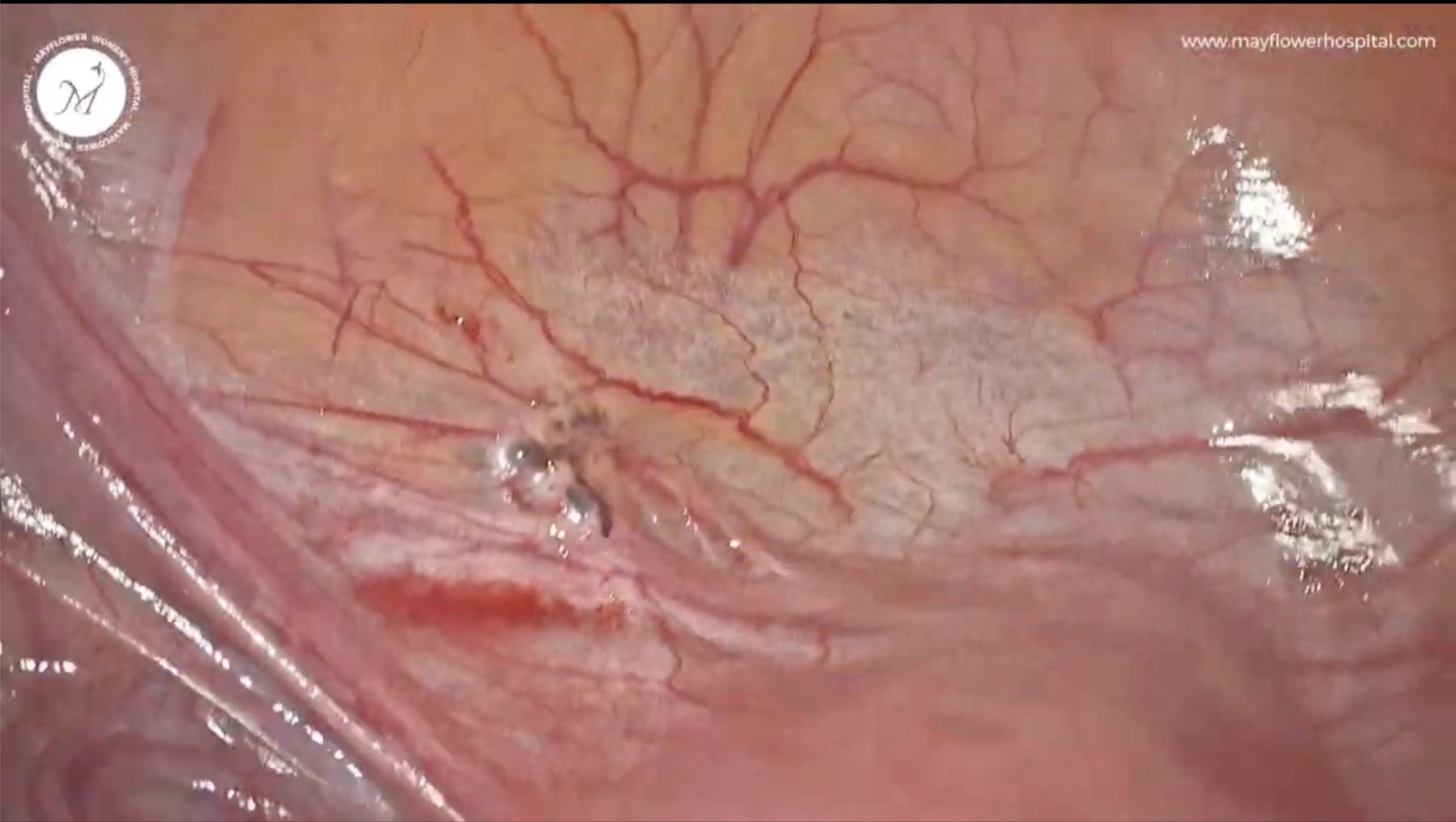
Bladder serosal nodule prior to excision.
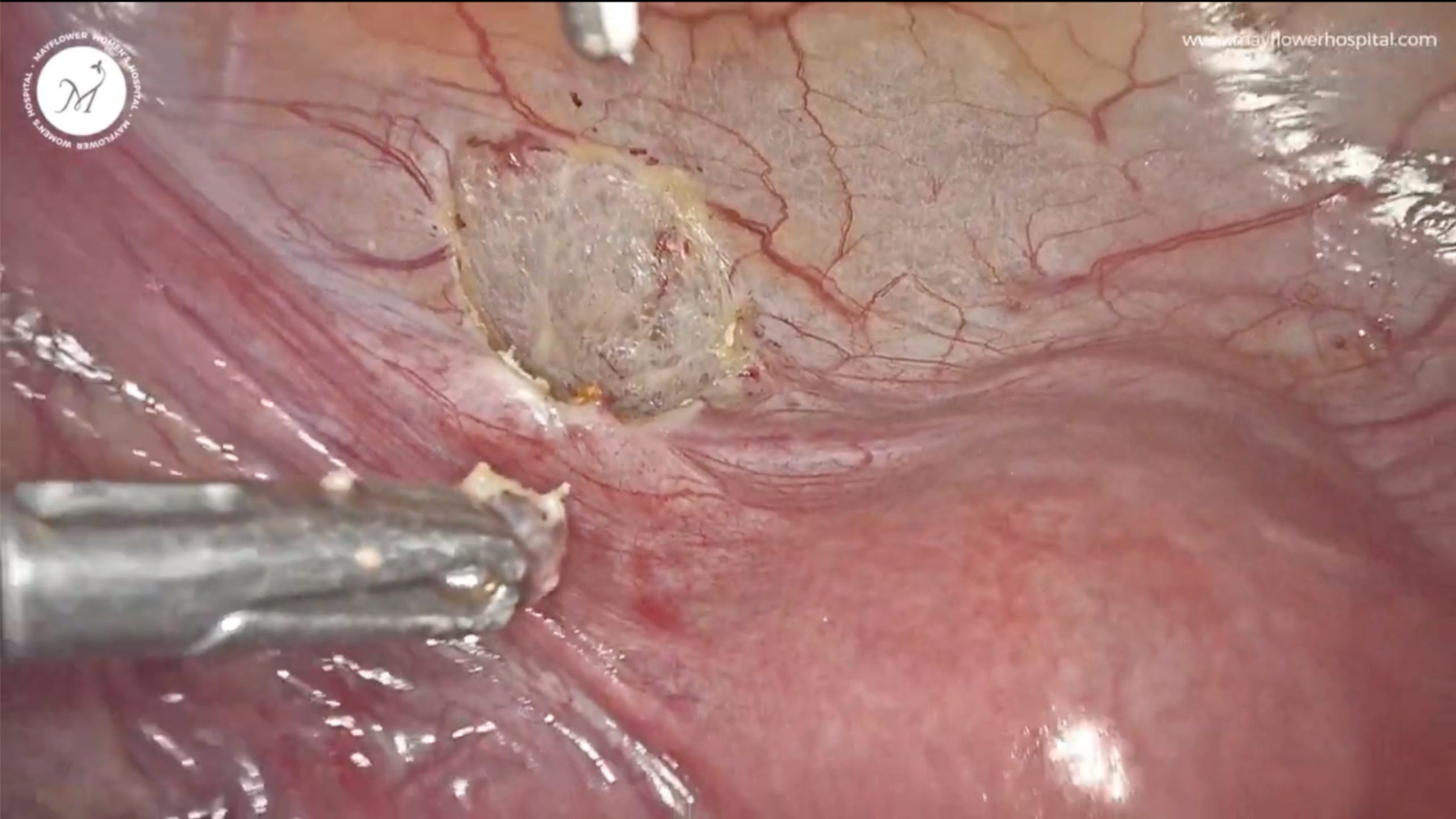
The same field after excision, with the bladder wall left intact.
With visible disease cleared, the fallopian tubes were assessed from the inside. Salpingoscopy — passing a scope through the natural tubal ostium to inspect the mucosal folds — grades tubal health on a standard 0-to-5 scale, from normal (0) to severely abnormal (5). Here, the right tube graded 0 and the left graded 1: both within a range favourable for spontaneous conception.
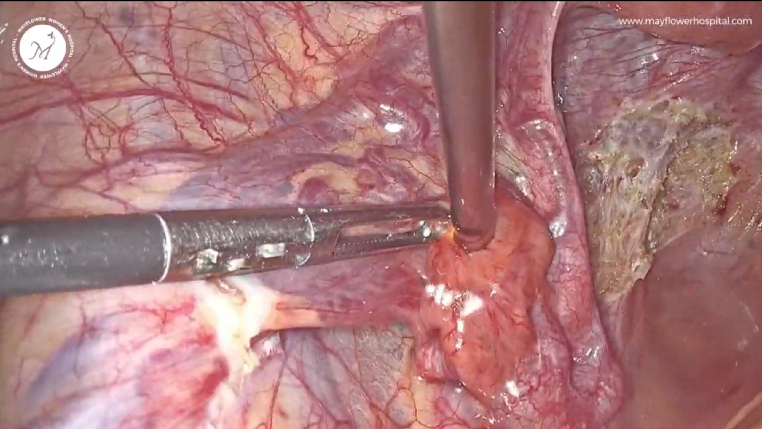
Entry into the natural orifice of the tube for endoluminal assessment.
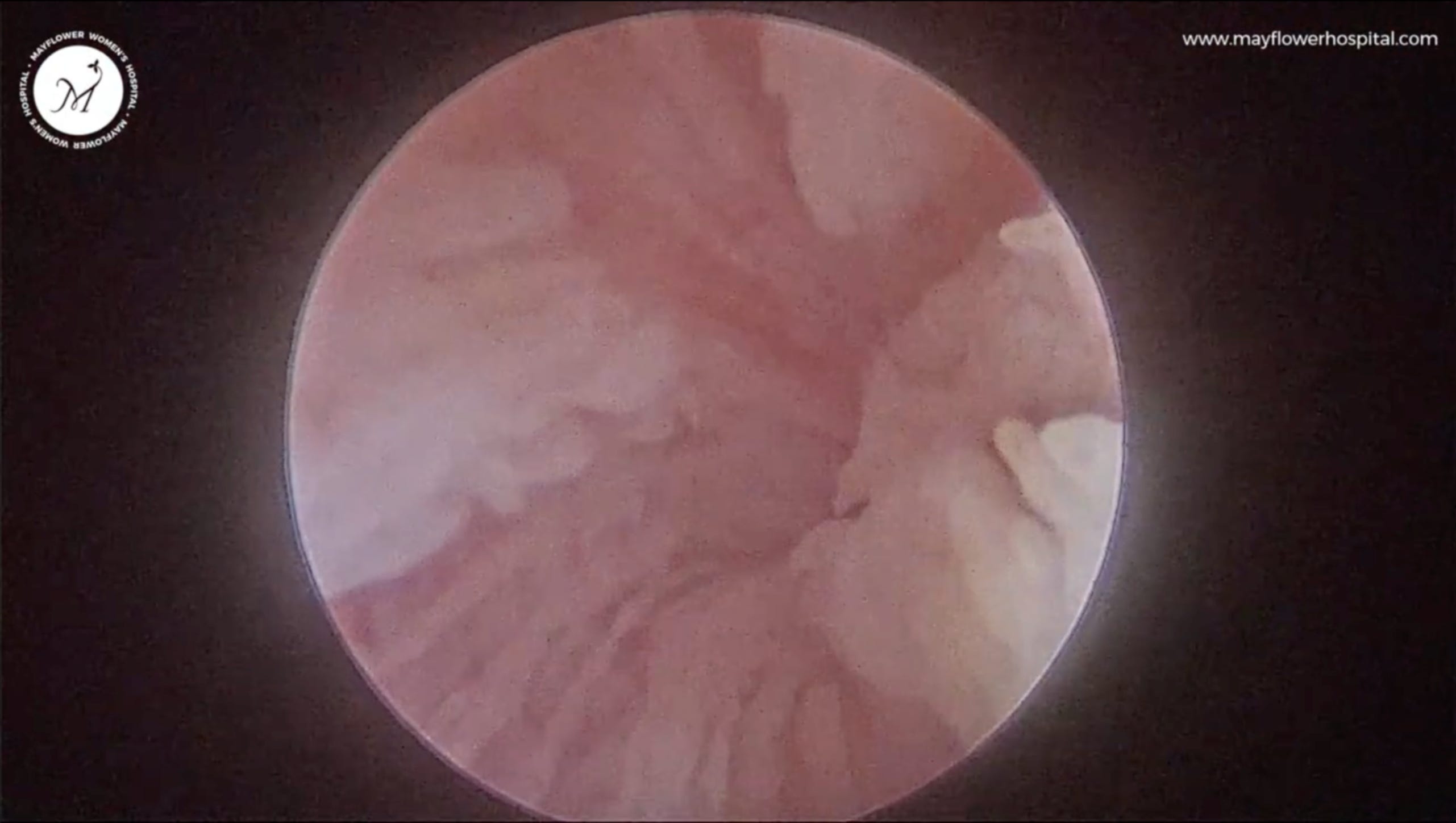
Internal view of the tubal mucosa. Both tubes graded favourably (right: 0, left: 1).
No further intervention was performed at this point. Consistent with the philosophy above — that minimally invasive surgery should mean minimal footprint, not maximal access — we did not proceed to additional tubal surgery or immediate ART referral. We advised the couple to attempt natural conception and followed her closely.
At six months, she was pregnant.
At caesarean section, we took the opportunity to inspect the posterior compartment directly. No adhesions. No endometriosis. The excision had held, and the pelvis behind the uterus looked as though nothing had ever been there.
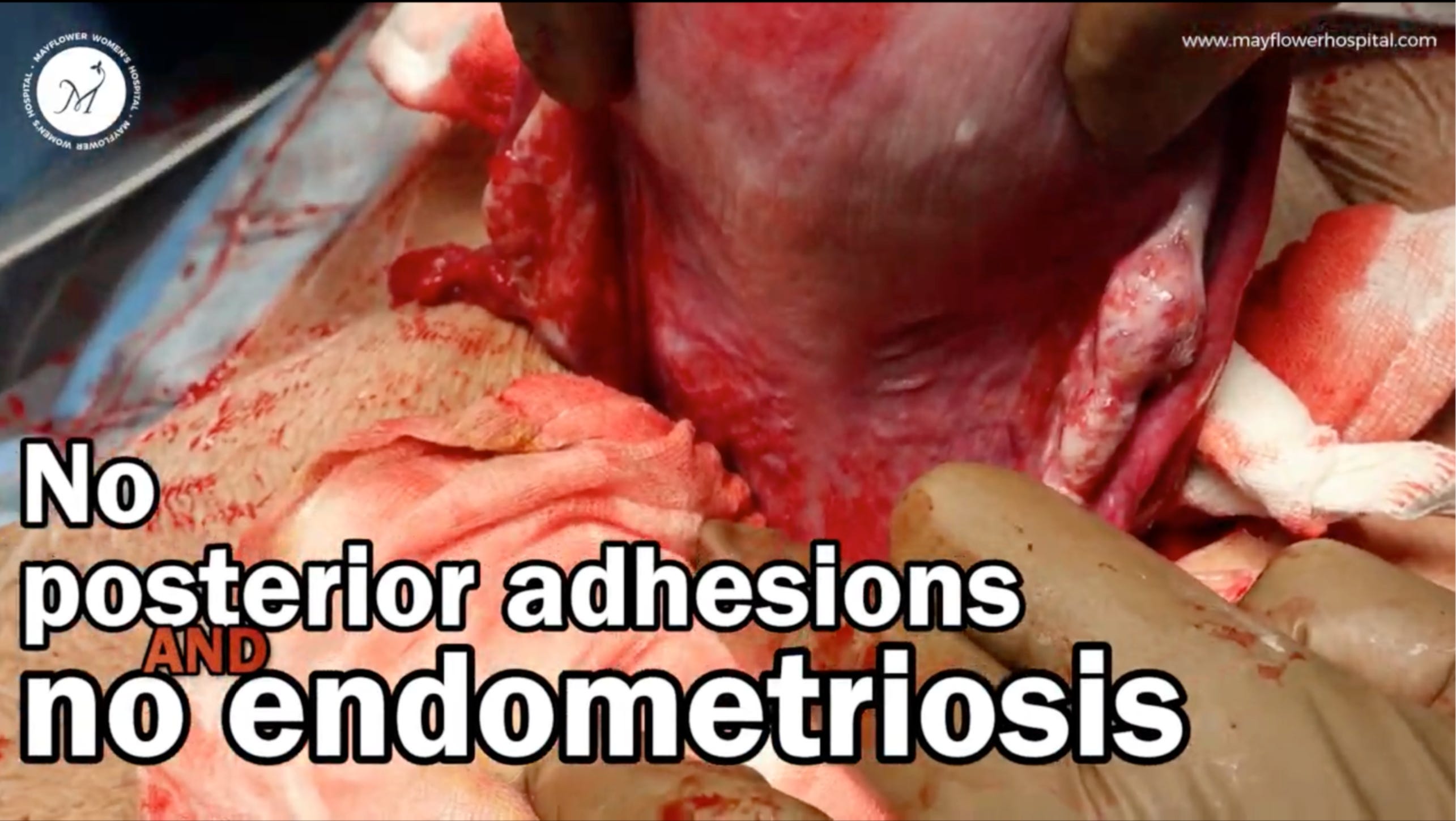
Posterior compartment at caesarean section: no adhesions, no residual disease.
It would have been easy to justify more surgery here. The disease was multifocal — parametrial, rectovaginal, vesical — and a maximalist would have found ample reason to extend the dissection: complete bilateral ureterolysis “for safety,” more aggressive tubal instrumentation, an earlier pivot to ART rather than a six-month wait.
None of it would have been indefensible. All of it would have been unnecessary.
Restraint, exercised at the right moments turned out to be as load-bearing to this pregnancy as anything we cut.
That’s it from us this week. See you in the next one.
No posts
Comments
Nothing yet. Say the first thing.
Sign in to join the conversation.