
There is a growing trend in laparoscopic gynaecology to explore the deep pelvis in endometriosis cases; even when the disease does not involve those structures. When disease is present, surgeons often adopt a maximally extensive approach: dissecting the retroperitoneum, isolating internal iliac vessels, identifying nerves, and exposing the base of the pelvis.
The question is worth asking: is access, in and of itself, the goal? Or should the extent of surgery be dictated entirely by the extent of disease?
This week’s case offers a different perspective, grounded in precision, restraint, and anatomical respect.
We present a case of endometriosis involving the lateral deep pelvis. This was not a plaque-type lesion; rather, it represented a peritoneal ingrowth extending into the deep pelvis, a kind of open-topped “deep well,” comparable in character to a peritoneal inclusion cyst but with peritoneal endometriosis involvement and an open apex. The depth and architecture of this lesion demanded a methodical, structured operative approach.
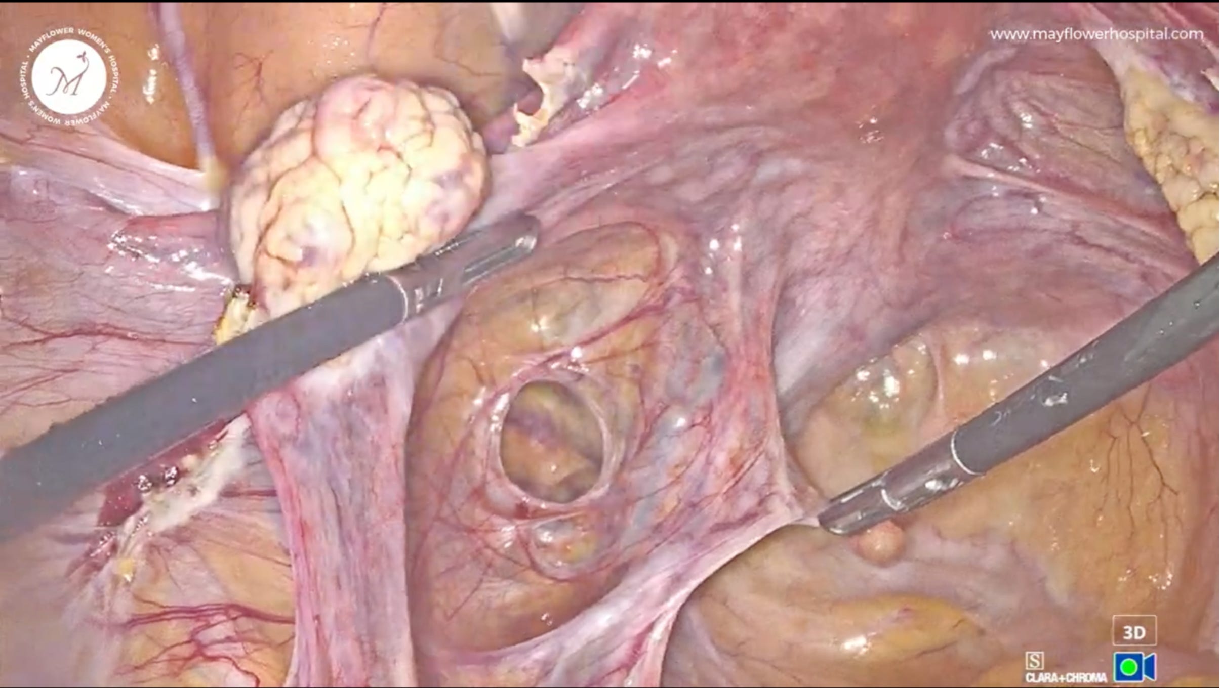
As with all cases of this nature, surgery began with ureterolysis at the pelvic brim. Dissection was performed on the anterior surface of the ureter using the Tenore device, utilising a combination of dissection, traction, counter-traction, and the cavitational effect unique to ultrasonic instrumentation.
The objectives of this step were two-fold:
Lateralise the ureter to create a safe working corridor
Enable complete excision of the diseased peritoneum without ureteric injury
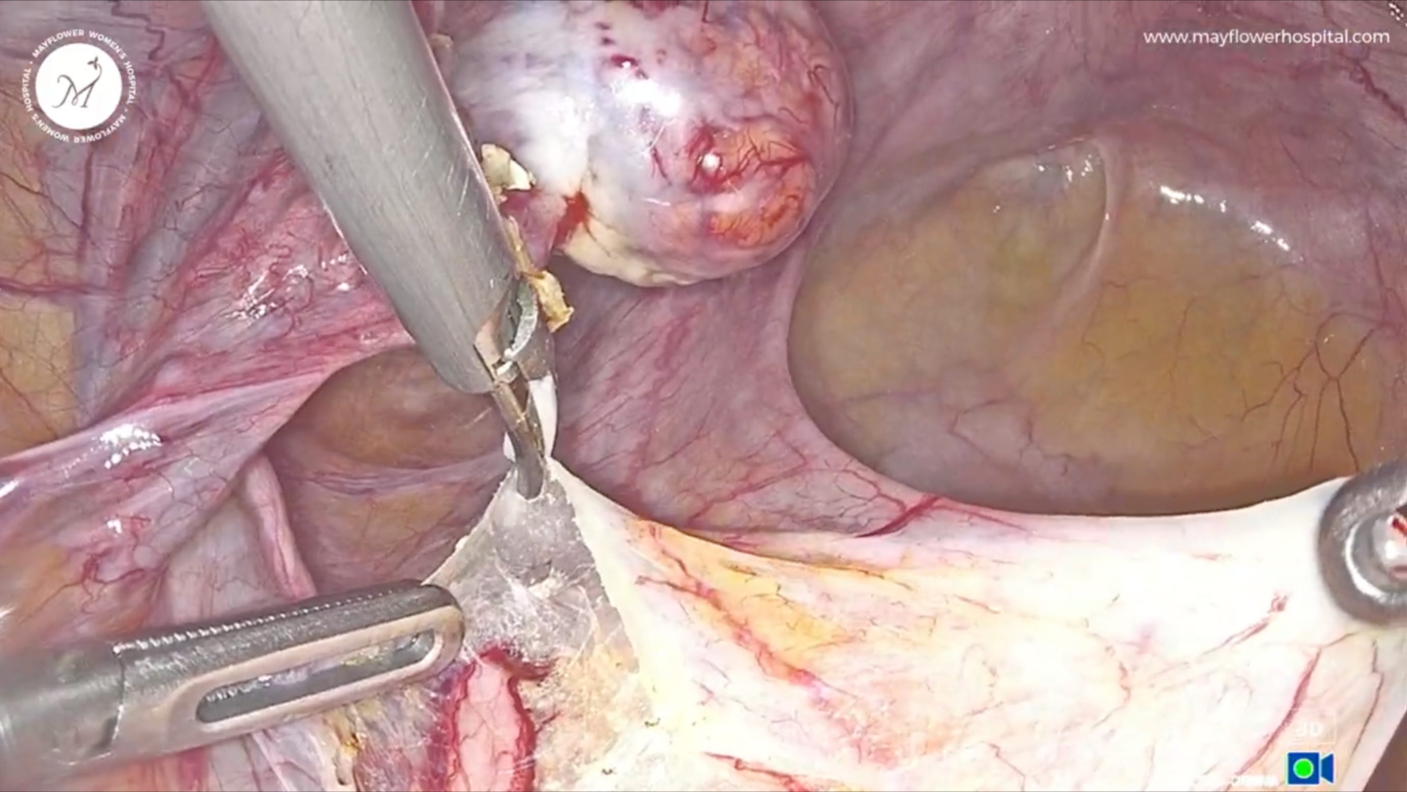
Attention was then turned to what we describe as the “upper wing of the butterfly” the pelvic brim peritoneum. At the pelvic brim, the dissected peritoneal leaflet was elevated with care taken to protect the infundibulopelvic (IP) ligament.
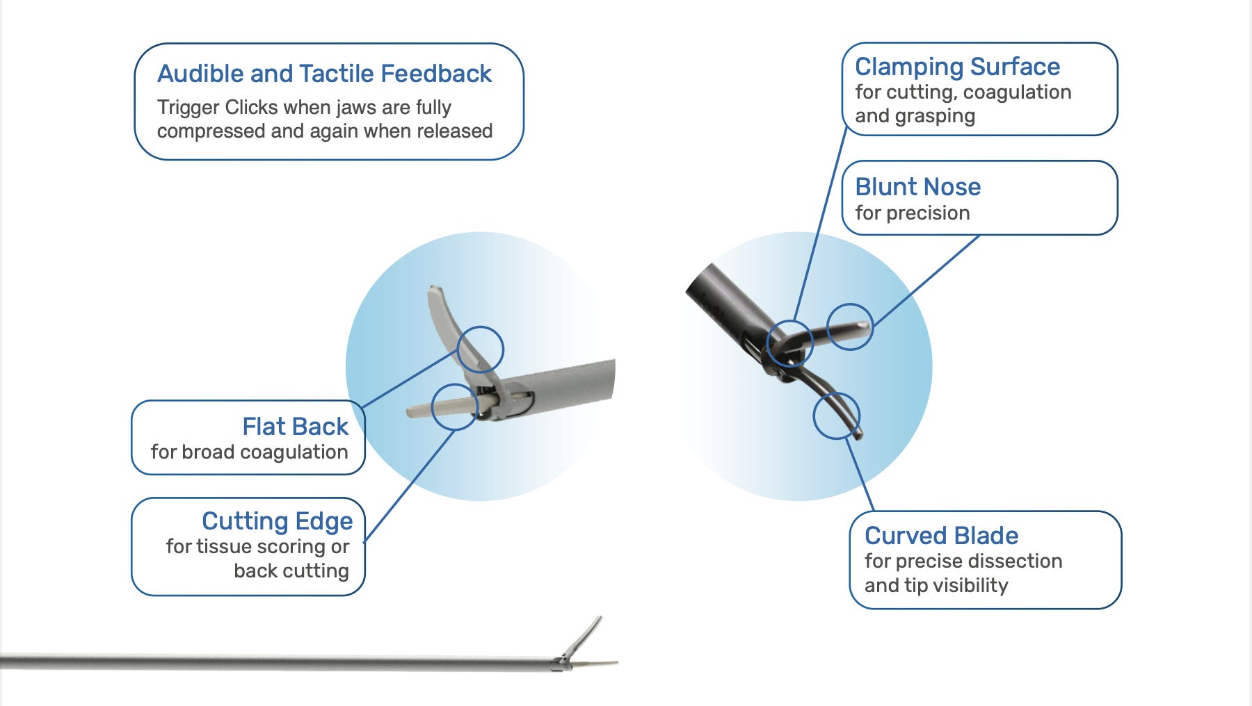
The Tenore device proved invaluable here: cavitational effect, dissection, and coagulation are all achieved through the mechanical movement of the jaw at a frequency of 55,000 displacements per second; a single instrument delivering precision and haemostasis simultaneously.
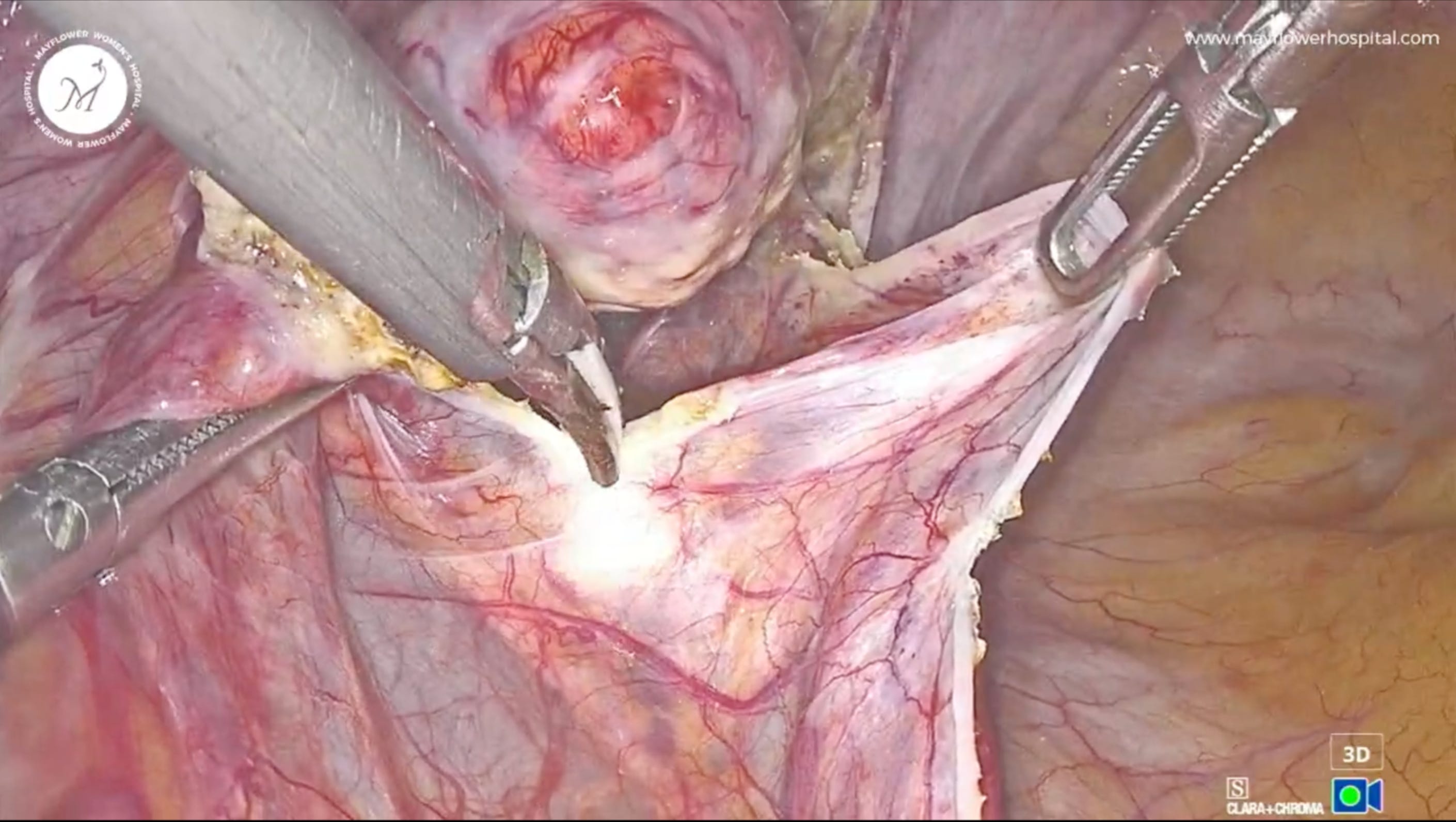
Peritoneum of the outer rim was systematically dissected and the dissection carried progressively deeper. As we moved into the depth of the lesion, a thorough knowledge of the surrounding anatomy became critical. The following structures were anticipated, and in some cases found to be distorted by disease:
Internal iliac artery
Uterine artery
Obliterated umbilical ligament
Vesical artery
Pudendal artery and accompanying veins
Sacral nerves
Given the anticipated anatomical distortion, a “predict and proceed” approach was adopted: before each activation of the Tenore jaw, the tissue was assessed tactilely and visually. “Smell the tissue” to confirm orientation. Only once the tissue plane was understood was the instrument activated. The goal at every step was to remove only the diseased peritoneum, leaving all named anatomical structures intact.
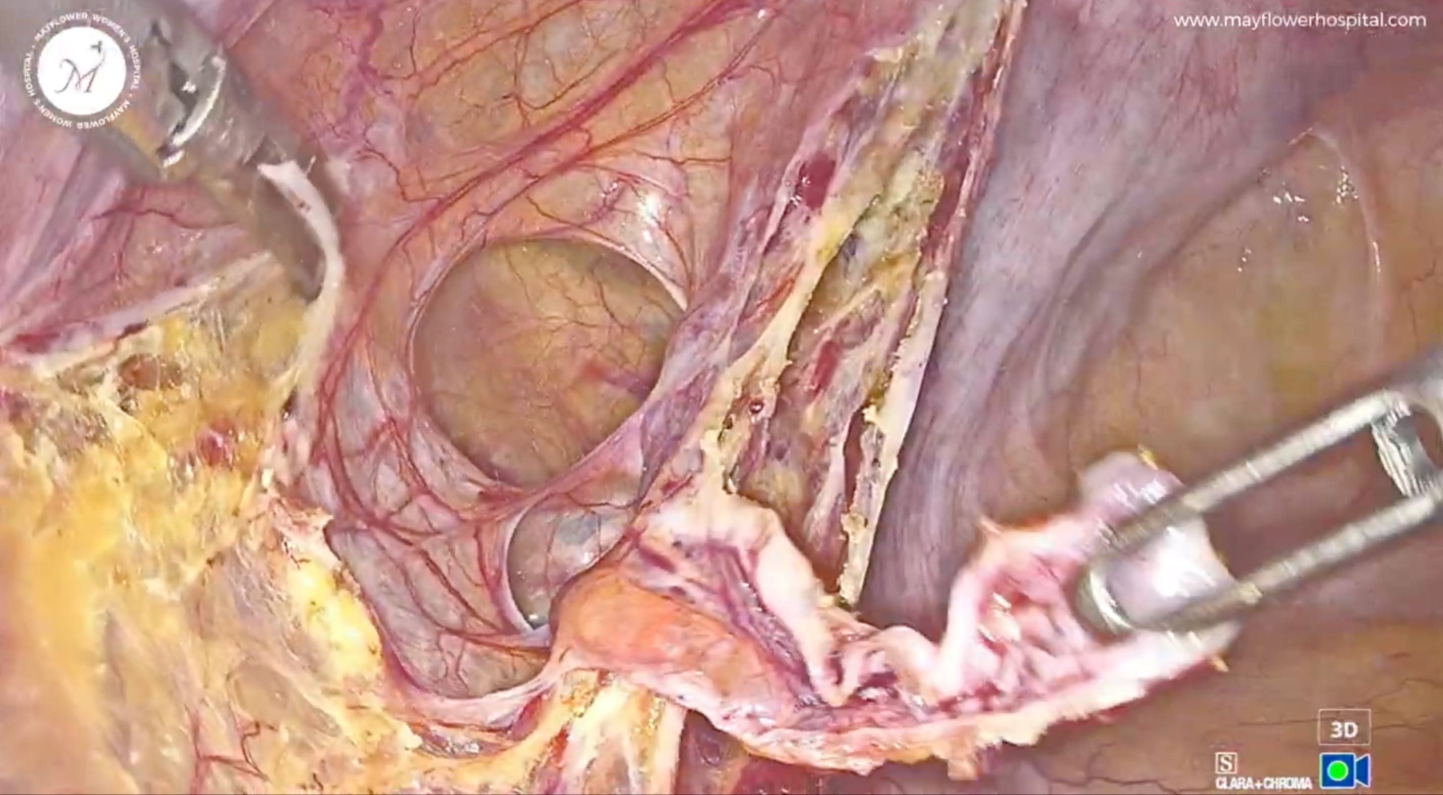
A secondary intraoperative review was performed to confirm the adequacy and safety of the excision. The assessment confirmed:
Depth of dissection achieved: Consistent with a complete removal
Quality of surgical excision: Satisfactory margins, no disease left behind
No injury to any vessels
Sacral nerves visualised and intact at the base of the pelvis
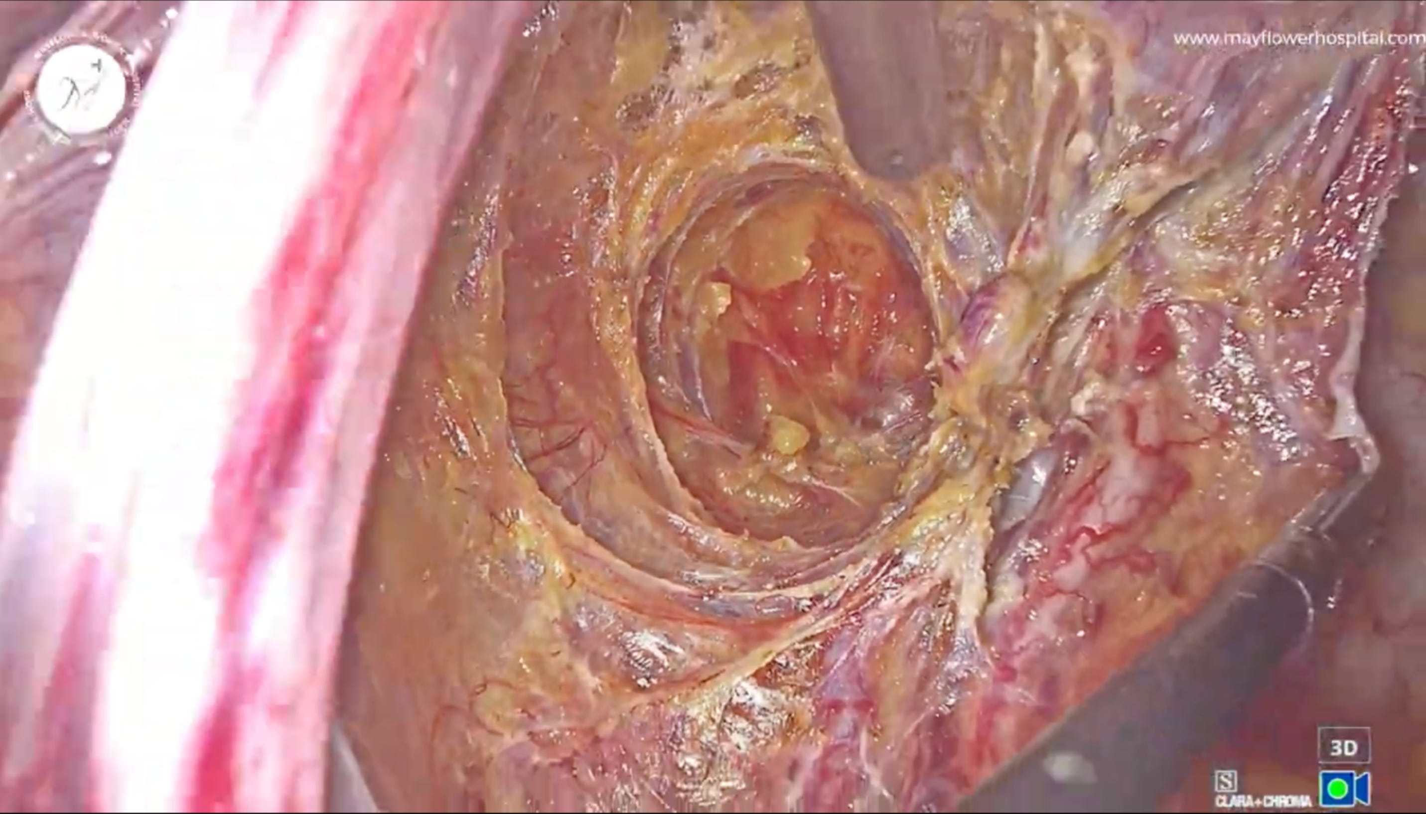
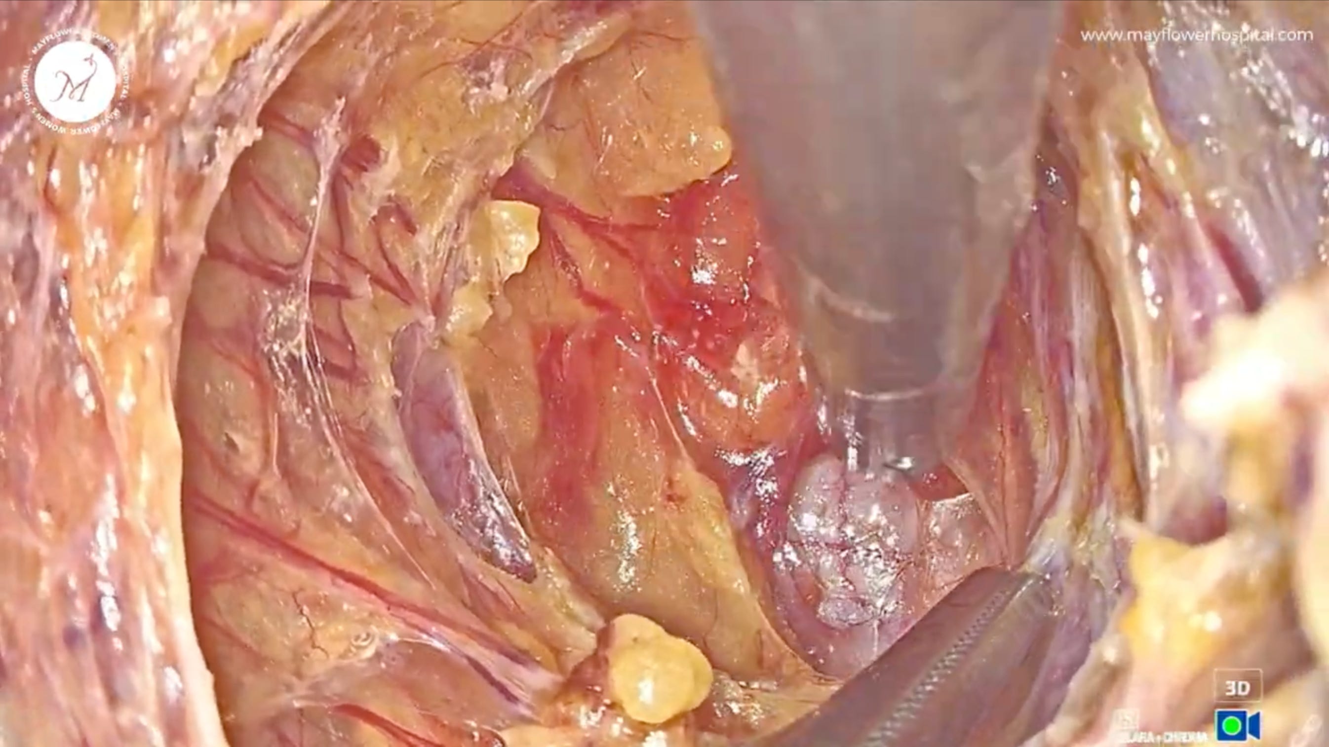
This case illustrates what truly minimally invasive surgery should mean. Minimal not just in access but also in collateral disruption. A tailored, conservative approach was employed: all anatomical structures preserved, no nerves damaged, and disease excised completely from the deepest recesses of the pelvis.
The Tenore device proved invaluable throughout delivering cavitational dissection, coagulation, and precise tissue handling within a single instrument. But technique and technology are secondary to surgical philosophy. The discipline of doing only what is necessary, informed at every step by anatomy, remains the cornerstone of safe and effective endometriosis surgery.
“Surgery need not be a demonstration. It need only be done well.”
That’s it from us this week. See you in the next one!
No posts
Comments
Nothing yet. Say the first thing.
Sign in to join the conversation.