
By Dr. David Traster, DC, MS, DACNB
Co-owner, The Neurologic Wellness Institute
Boca Raton • Chicago • Waukesha • Wood Dale
www.neurologicwellnessinstitute.com
The autonomic nervous system is often thought of as the body’s “automatic” control system, regulating heart rate, blood pressure, respiration, digestion, temperature, pupil size, immune function, and countless other physiological processes without conscious effort. Yet the autonomic nervous system is far more sophisticated than simply balancing sympathetic and parasympathetic activity. It represents an enormous network extending from peripheral receptors throughout the body to highly integrated circuits within the spinal cord, brainstem, cerebellum, hypothalamus, limbic system, and cerebral cortex. Every moment these structures continuously calculate how much blood pressure should rise, how rapidly the heart should beat, how much blood should be directed to skeletal muscle, skin, kidneys, or digestive organs, and how the body should prepare for physical or emotional stress.
Among the simplest yet most informative bedside autonomic tests is the isometric hand grip test. Although deceptively simple, this test challenges one of the most fundamental reflexes within the autonomic nervous system: the ability to generate sustained sympathetic activation while maintaining cardiovascular stability. When interpreted correctly, the hand grip test provides valuable insight into the integrity of both peripheral autonomic pathways and the central neural networks responsible for cardiovascular regulation.
The autonomic nervous system consists of peripheral and central components working together as a single integrated network. The peripheral autonomic nervous system includes sympathetic ganglia, parasympathetic ganglia, postganglionic nerve fibers, autonomic plexuses, baroreceptors located within the carotid sinus and aortic arch, cardiopulmonary stretch receptors, chemoreceptors, visceral sensory receptors, adrenal medulla, enteric nervous system, and numerous autonomic receptors located throughout nearly every organ. These peripheral sensors continuously report changes in blood pressure, blood chemistry, oxygen levels, carbon dioxide concentration, tissue stretch, inflammation, and organ function back to the central nervous system.
The central autonomic nervous system is considerably more complex. Cardiovascular regulation depends upon continuous communication among the nucleus tractus solitarius (NTS), dorsal motor nucleus of the vagus, nucleus ambiguus, caudal ventrolateral medulla (CVLM), rostral ventrolateral medulla (RVLM), parabrachial nucleus, periaqueductal gray, hypothalamus, cerebellar vermis, fastigial nucleus, vestibular nuclei, insular cortex, anterior cingulate cortex, medial prefrontal cortex, amygdala, hippocampus, and multiple regions of the reticular formation. Rather than functioning independently, these regions constantly exchange information to determine whether sympathetic activity should increase or decrease.
At the center of sympathetic cardiovascular control lies the intermediolateral cell column (IML) of the spinal cord. Located primarily between spinal levels T1 through L2, the IML contains the cell bodies of preganglionic sympathetic neurons. These neurons represent the final common pathway through which the brain increases vascular tone, heart rate, myocardial contractility, adrenal activation, sweating, and numerous other sympathetic functions.
Whenever blood pressure needs to rise, descending signals from the brain activate neurons within the IML. These neurons then project to sympathetic chain ganglia, where postganglionic neurons ultimately innervate blood vessels, the heart, adrenal glands, kidneys, and other organs. Without proper activation of the IML, the body cannot adequately increase systemic vascular resistance or maintain blood pressure during physical or emotional stress.
Multiple regions of the brain increase sympathetic outflow by exciting neurons within the intermediolateral cell column. Perhaps the most important cardiovascular excitatory center is the rostral ventrolateral medulla (RVLM). The RVLM provides continuous tonic excitation to sympathetic preganglionic neurons and largely determines resting sympathetic vascular tone. Even at rest, the RVLM maintains sufficient activity to preserve normal arterial blood pressure.
The hypothalamus provides powerful descending sympathetic drive during exercise, thermal stress, dehydration, emotional arousal, fear, pain, and metabolic demands. The periaqueductal gray coordinates autonomic responses associated with defensive behaviors and pain modulation. The amygdala activates sympathetic pathways during emotional stress, anxiety, fear, and threat perception. The insular cortex—particularly the right insula—plays a major role in cardiovascular sympathetic regulation. The anterior cingulate cortex contributes to autonomic responses during effort, attention, and pain.
Motor cortex also contributes through a mechanism known as central command, whereby voluntary movement simultaneously activates skeletal muscle and sympathetic cardiovascular centers before exercise even begins. Vestibular nuclei excite sympathetic pathways through the vestibulosympathetic reflex, allowing blood pressure to rise appropriately during changes in head position and posture. The cerebellar fastigial nucleus modulates sympathetic output during postural adjustments and cardiovascular challenges. Together these structures converge upon the RVLM and spinal sympathetic neurons, allowing rapid cardiovascular adaptation.
Sympathetic activation must also be restrained. Excessive sympathetic activity is metabolically costly and can damage blood vessels, the heart, kidneys, and brain. The most important inhibitory center is the caudal ventrolateral medulla (CVLM). The CVLM receives excitatory input from the nucleus tractus solitarius after baroreceptor activation. It then inhibits the RVLM using GABAergic neurons, thereby decreasing excitation reaching the IML.
The nucleus tractus solitarius (NTS) serves as the primary sensory integration center for baroreceptor, chemoreceptor, pulmonary stretch receptor, and visceral afferent information. Increased blood pressure activates carotid sinus and aortic arch baroreceptors, which excite the NTS. The NTS subsequently activates the CVLM, suppressing RVLM activity and reducing sympathetic output. Parasympathetic nuclei including the nucleus ambiguus and dorsal motor nucleus of the vagus simultaneously increase vagal tone to slow heart rate.
Higher cortical structures such as the medial prefrontal cortex also exert inhibitory control over hypothalamic and limbic sympathetic centers, helping regulate emotional autonomic responses. The cerebellum further refines autonomic responses by preventing excessive sympathetic activation during movement and postural adjustments. The balance between excitatory and inhibitory pathways ultimately determines the amount of sympathetic activity leaving the spinal cord.
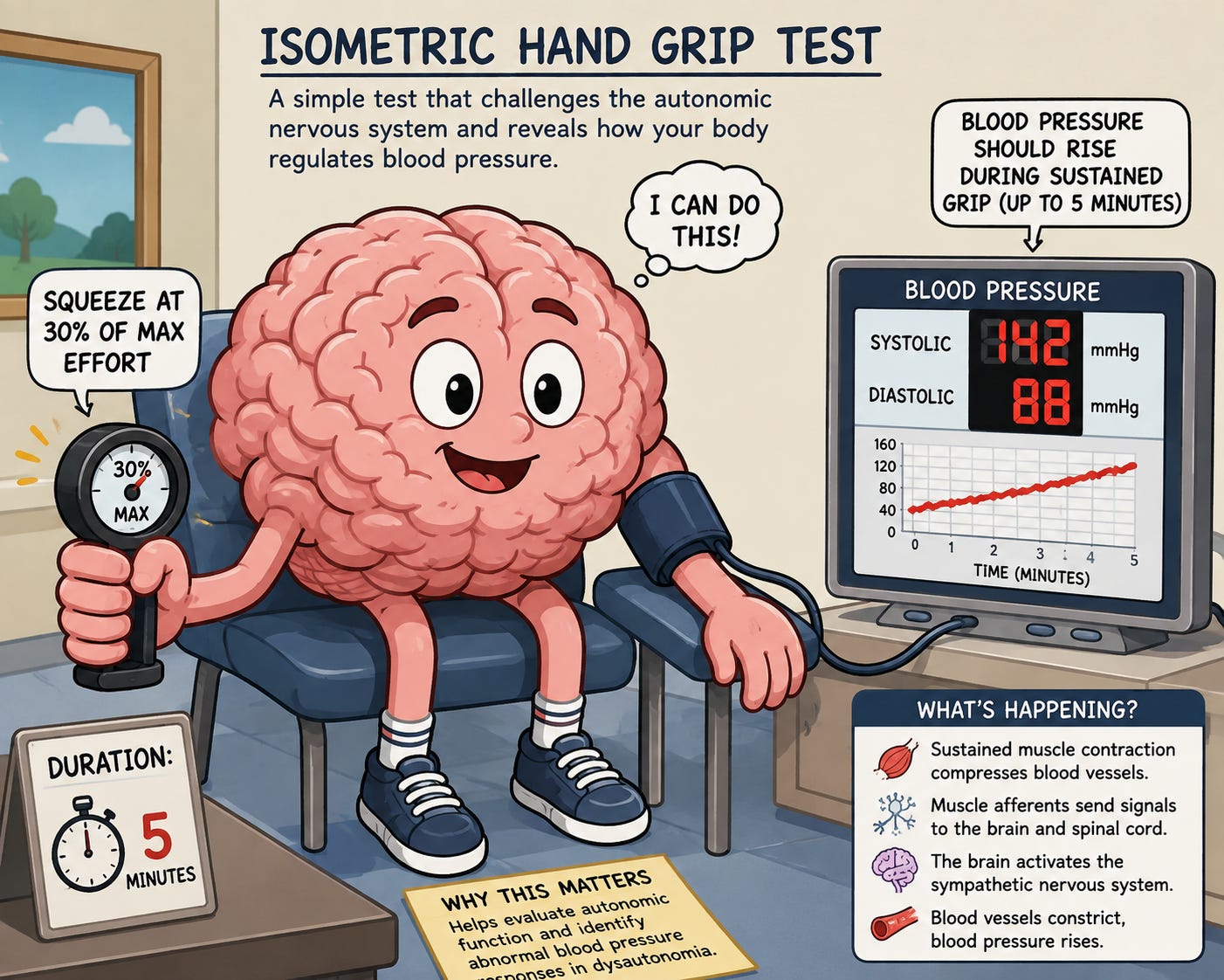
The isometric hand grip test appears deceptively simple. The patient squeezes a dynamometer or grip device at approximately 30% of maximal voluntary contraction for up to five minutes. Unlike dynamic exercise, isometric contraction produces sustained compression of intramuscular blood vessels. As contraction continues, local blood flow becomes progressively restricted. This leads to accumulation of metabolites including lactate, hydrogen ions, potassium, adenosine, prostaglandins, ATP, and bradykinin. These metabolites activate group III and group IV skeletal muscle afferents, collectively known as the exercise pressor reflex.
These afferents ascend into the spinal cord before projecting to the nucleus tractus solitarius, parabrachial nucleus, hypothalamus, and medullary cardiovascular centers. Simultaneously, voluntary effort activates motor cortex, which generates central command signals that independently stimulate sympathetic centers before metabolic accumulation even occurs.
The combined effects of central command and the exercise pressor reflex produce progressive sympathetic activation. Descending excitation increases activity within the RVLM and ultimately the intermediolateral cell column. Sympathetic nerves release norepinephrine onto resistance vessels throughout the body, producing systemic vasoconstriction and increasing total peripheral resistance. Cardiac sympathetic fibers increase contractility and often modestly increase heart rate. The adrenal medulla may also release epinephrine.
In healthy individuals, systolic blood pressure typically rises, but the hallmark response is a sustained increase in diastolic blood pressure, generally by at least 16 mmHg and often 20 mmHg or more during the latter stages of the test. This reflects increased peripheral vascular resistance rather than simply increased cardiac output.
Patients with dysautonomia often demonstrate abnormal responses because one or more portions of the autonomic network fail to generate or regulate appropriate sympathetic activation. The pattern of abnormality can provide important physiological clues.
When blood pressure fails to increase appropriately, several mechanisms should be considered. Peripheral sympathetic neuropathy may prevent adequate neurotransmitter release despite intact central activation. Damage to preganglionic neurons within the intermediolateral cell column may impair sympathetic output. Degeneration of sympathetic ganglia or postganglionic fibers can reduce vasoconstriction. Impaired RVLM function may fail to generate sufficient descending sympathetic drive.
Reduced hypothalamic activation, impaired exercise pressor reflexes, abnormalities of muscle afferent signaling, spinal cord disease, neurodegenerative disorders such as multiple system atrophy, diabetic autonomic neuropathy, autoimmune autonomic ganglionopathy, advanced Parkinsonian syndromes, or severe peripheral neuropathies may all contribute. Clinically, these patients often experience exercise intolerance, orthostatic hypotension, fatigue, dizziness, reduced vascular resistance, and difficulty maintaining blood pressure during stress.
An exaggerated blood pressure response suggests excessive sympathetic activation or inadequate inhibitory control. Several central mechanisms may contribute. Reduced baroreflex sensitivity prevents normal buffering of sympathetic activity. The nucleus tractus solitarius may inadequately activate the CVLM. The CVLM may fail to sufficiently inhibit the RVLM. The RVLM itself may become hyperexcitable. Loss of inhibitory cortical regulation from the medial prefrontal cortex or excessive activation from the amygdala and hypothalamus can further amplify sympathetic output.
Patients with hyperadrenergic postural orthostatic tachycardia syndrome, chronic anxiety disorders, chronic pain syndromes, autonomic hypersensitivity, some forms of traumatic brain injury, and certain neurodegenerative conditions may demonstrate excessive responses. Clinically these patients often report palpitations, tremor, headaches, flushing, elevated norepinephrine levels, cold extremities from excessive vasoconstriction, episodic hypertension, and exercise intolerance despite apparently robust sympathetic activation.
Perhaps the most concerning abnormal response occurs when blood pressure actually decreases during sustained isometric contraction. This suggests a profound failure of sympathetic cardiovascular compensation.
Potential mechanisms include severe autonomic neuropathy, failure of RVLM activation, spinal cord lesions affecting the intermediolateral cell column, advanced neurodegeneration, impaired descending hypothalamic pathways, severe baroreflex dysfunction, or paradoxical vasodilatory responses.
In some patients, excessive vagal activation or abnormal central autonomic integration may override the expected sympathetic response. A falling blood pressure during sustained muscular effort indicates that the nervous system cannot maintain vascular resistance when metabolic demand increases. These patients frequently experience severe orthostatic intolerance, syncope, exercise-induced hypotension, and marked fatigue because the central autonomic network cannot adequately support cardiovascular homeostasis.
The isometric hand grip test should never be viewed as simply a blood pressure measurement. It represents a functional challenge to the entire autonomic nervous system. Every phase of the response depends upon healthy skeletal muscle afferents, intact peripheral autonomic nerves, properly functioning sympathetic ganglia, responsive vascular smooth muscle, and an extraordinarily complex network of brainstem, cerebellar, hypothalamic, limbic, cortical, and spinal circuits.
An abnormal response does not immediately identify a single lesion. Instead, it reveals that one or more components of the central autonomic network have failed to appropriately generate, modulate, or execute sympathetic cardiovascular control. When interpreted alongside tilt testing, the Valsalva maneuver, heart rate variability, deep breathing, sudomotor testing, vestibular evaluation, and detailed neurological examination, the isometric hand grip test becomes an invaluable tool for understanding where autonomic regulation may be breaking down.
Rather than serving as merely another cardiovascular stress test, the isometric hand grip test offers clinicians a dynamic glimpse into the integrity of one of the body’s most sophisticated control systems. Every rise—or failure to rise—in blood pressure reflects the coordinated activity of an immense neural network that continuously works to keep cerebral perfusion stable, maintain organ function, and preserve homeostasis in the face of ever-changing physiological demands.
Benarroch, E. E. (2012). The central autonomic network: Functional organization, dysfunction, and perspective. Mayo Clinic Proceedings, 87(10), 988–1001.
Freeman, R., Wieling, W., Axelrod, F. B., Benditt, D. G., Benarroch, E. E., Biaggioni, I., Cheshire, W. P., Chelimsky, T., Cortelli, P., Gibbons, C. H., Goldstein, D. S., Hainsworth, R., Hilz, M. J., Jacob, G., Kaufmann, H., Jordan, J., Lipsitz, L. A., Levine, B. D., Low, P. A., Mathias, C. J., Raj, S. R., Robertson, D., Sandroni, P., Schatz, I., Schondorff, R., Stewart, J. M., van Dijk, J. G., & Consensus Committee of the American Autonomic Society and the American Academy of Neurology. (2011). Consensus statement on the definition of orthostatic hypotension, neurally mediated syncope, and the autonomic disorders. Clinical Autonomic Research, 21(2), 69–72.
Hall, J. E. (2021). Guyton and Hall textbook of medical physiology (14th ed.). Elsevier.
Low, P. A. (Ed.). (2008). Clinical autonomic disorders: Evaluation and management (3rd ed.). Lippincott Williams & Wilkins.
Victor, R. G., Seals, D. R., & Mark, A. L. (1987). Differential control of heart rate and sympathetic nerve activity during dynamic exercise and static exercise in humans. Journal of Clinical Investigation, 79(2), 508–516.
No posts
Comments
Nothing yet. Say the first thing.
Sign in to join the conversation.