The rise of artificial intelligence in medicine is reshaping how orthopedic surgeons diagnose, plan, and deliver care. From robotics in the operating room to AI-driven diagnostics and bone segmentation tools, orthopedic innovation is advancing rapidly. Yet, adoption in everyday surgical practice has been more measured. For example, in 2022, robot-assisted technology accounted for 11.6% of total…
The integration of cone beam CT imaging in research labs is revolutionizing orthopedic investigations. Dr. Zong-Ming Li, PhD, David Jordan, and Trevour Greene from the University of Arizona discuss the transformative impact of the InReach CT system from CurveBeam AI on their work. The Hand Research Laboratory at the University of Arizona focuses on solving orthopedic-related clinical problems,…
Rapid advancements in surgical planning techniques are propelling the field of orthopedic technology into a transformative phase. João Pedro Ribeiro, the Chairman and CEO of PeekMed, is at the forefront of this change. His story, from a biomedical engineer to an industry leader, highlights the impact of innovation in healthcare. As we enter an era where technology and medicine intersect more than…
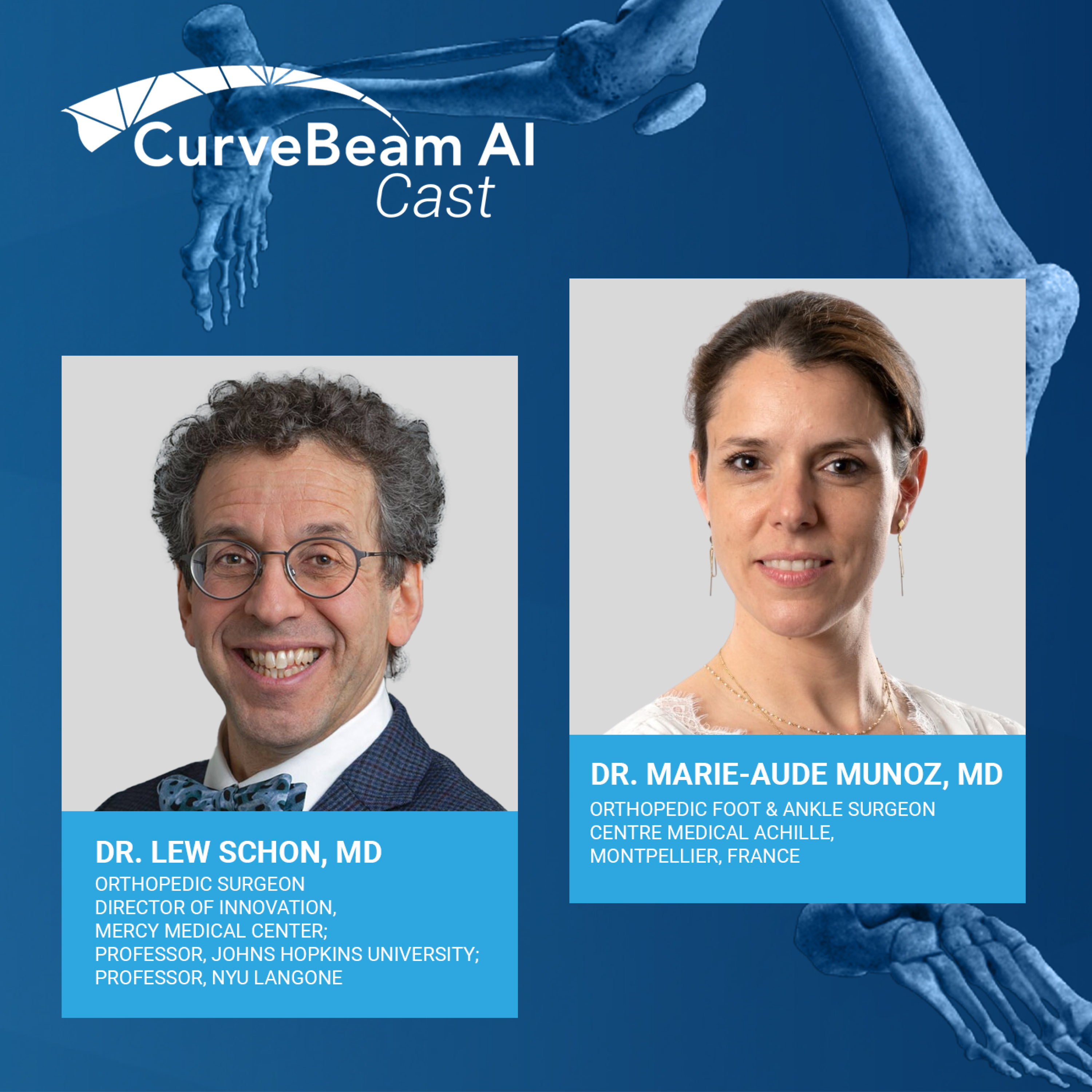
In the latest CurveBeam AI Cast episode, recorded at the AOFAS Annual Meeting 2023, host Dr. Lew Schon , MD, Director of Innovation at Mercy Medical Center and Professor at John Hopkins University and NYU Langone , engages in a riveting conversation with Dr. Marie-Aude Munoz, MD , a distinguished Orthopedic Foot & Ankle Surgeon from Montpellier, France. Together, they explore Dr. Munoz's…
The introduction of weight-bearing CT scans has revolutionized how surgeons assess and treat various orthopedic conditions. Research indicates this technology can provide clinicians with in-depth, real-time insights, allowing for better patient outcomes and more accurate diagnoses. How is weight bearing CT technology influencing the approach of orthopedic surgeons, and how are patients benefitting…
The benefits of WBCT (Weight Bearing Computed Tomography) usher in a new era of orthopedic diagnostics, illuminating previously obscure aspects of hindfoot alignment. The shift from 2D X-ray technology to 3D imaging has been profound, offering a more intricate understanding of bone structures and alignments, which is critical for effective treatment plans. Numerous research articles point to the…
In recent years, the orthopedics field has been gravitating towards using 3D imaging technologies to foster a deeper understanding of bone structures and alignments. A notable driver of this shift is the application of Weight bearing Computed Tomography (WBCT), which has emerged as a game-changing tool for evaluating hindfoot alignment. What might the integration of weight bearing CT signify for…
Axiometrics may be new but it’s set on revolutionizing orthopedics forever. Every year, approximately two million Americans suffer from acute ankle sprains, and bone and joint injuries in the lower limbs. While most of these injuries heal with time, there is a subset of patients who experience chronic ankle instability, enduring persistent pain and limited mobility. Unfortunately, the underlying…
Imaging and diagnostics are important facets in healthcare and nearly every area of medicine relies on this technology to diagnose or monitor conditions. In orthopedics, it is an especially vital tool, as it allows surgeons to discover and perform successful procedures for their patients. Currently, improved AI imaging technology is helping to support orthopedic surgeons in identifying and better…
The foot and ankle are complex structures comprised of 26 small bones and 29 muscles. With so many structures and an incredible amount of variation in how deformities can present, surgical corrections aren’t so simple. For example, there are more than 100 operations that can be performed for hallux valgus deformities, commonly known as bunions. 3D planning helps to address the variations and…