
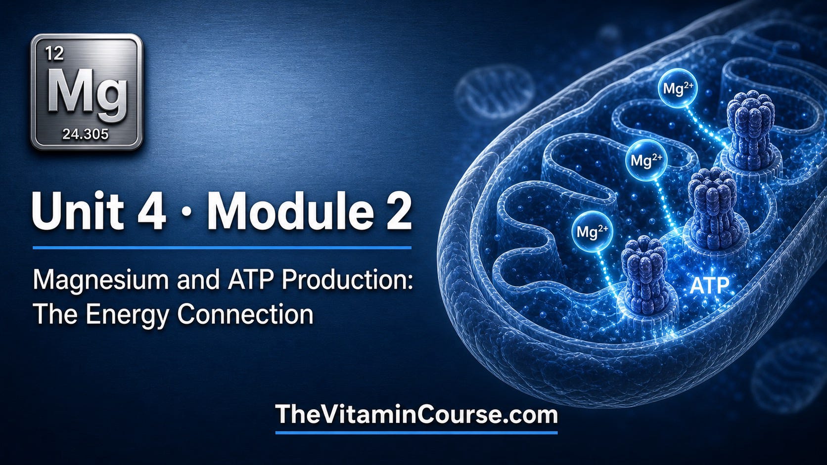
Welcome to Module 4.2 of Magnesium Mastery. In the previous module, we established the broad scope of magnesium’s role in cellular and metabolic function. In this module, we focus on the most foundational of those roles: magnesium’s indispensable position at the center of ATP production and bioenergetics. Without understanding this relationship, it is difficult to fully appreciate why magnesium deficiency can produce such a wide and seemingly unrelated constellation of symptoms. By the end of this module, you will understand exactly how magnesium enables the cell to produce, transfer, and utilize energy and what happens at the molecular and clinical level when it does not.
This article continues the building of a comprehensive course on magnesium which will be part of a much larger project: a free, fully open-access mega-course covering all vitamins and minerals.
If it has been a while since you’ve followed the series, or if you are new, here are the previous units to review:
Unit 1: Introduction to Magnesium
Unit 2: Magnesium and the Cardiovascular System
Unit 3: Magnesium and the Endocrine System
All of these units will eventually be organized into structured free courses hosted on a dedicated platform (TheVitaminCourse.com), which is currently under development. The long-term goal is to provide high-quality, comprehensive education—completely free—to professionals, laypeople, and even younger learners.
This work is offered freely as part of a larger mission. If you would like to support and help sustain this effort, maintaining a paid subscription is one of the most direct ways to do so and enables continued research, writing, and course development.
Every cell in the human body runs on adenosine triphosphate (ATP), the universal energy currency of life. Students and clinicians alike are taught that ATP provides the energy for muscle contraction, ion transport, biosynthesis, and signaling. What is far less commonly taught is that free ATP, on its own, is not the biologically active form.
To be used as a substrate by the vast majority of ATP-dependent enzymes, ATP must first bind a magnesium ion. The resulting complex, Mg-ATP, is the true substrate for hundreds of enzymatic reactions, including those central to mitochondrial energy metabolism. The magnesium ion coordinates with the beta and gamma phosphate groups of ATP, stabilizing the molecule’s highly charged triphosphate tail and positioning it correctly within enzyme active sites. Without this coordination, ATP is geometrically and electrostatically unable to engage the catalytic machinery of most ATP-dependent enzymes efficiently. [1, 2, 3]
This distinction has profound clinical implications. A patient may have a serum ATP level that appears adequate and yet still be functionally energy-depleted at the cellular level if intracellular free magnesium is insufficient to generate adequate Mg-ATP. The cell’s energetic problem in magnesium deficiency is not simply that ATP is made in lower quantities, it is that the ATP available cannot be fully utilized. [2, 4, 5]
The specificity of the Mg-ATP requirement extends across metabolic pathways. Hexokinase, phosphofructokinase, pyruvate kinase, and adenylate kinase, all key enzymes in glycolysis, use Mg-ATP as their substrate. ATP synthase, the enzyme responsible for producing the majority of cellular ATP via mitochondrial oxidative phosphorylation, requires magnesium both as a substrate component and as a direct activator. This means magnesium sits at both ends of the energy cycle: it is needed to make ATP via the mitochondrial machinery, and it is needed to use ATP once it has been made. [3, 6]
Mitochondria are not passive recipients of cytosolic magnesium. They maintain their own regulated pool of Mg²⁺, and the dynamics of this intramitochondrial magnesium are increasingly recognized as critical determinants of cellular energy status and metabolic resilience. [4, 7]
The primary gateway for magnesium entry into the mitochondrial matrix is MRS2 (Mitochondria RNA Splicing 2 protein), a magnesium-selective channel embedded in the inner mitochondrial membrane. MRS2 is the mammalian homolog of a yeast splicing factor that was found to be structurally similar to bacterial CorA magnesium channels. [4, 8]
Landmark research using fluorescence imaging and metabolomic analysis in MRS2 knockdown cells demonstrated that disruption of mitochondrial Mg²⁺ homeostasis fundamentally alters energy metabolism. Cells with reduced MRS2 activity showed: [4, 8]
A shift in mitochondrial energy metabolism away from oxidative phosphorylation
Altered mitochondrial morphology consistent with dysfunction
Reduced ATP production
Increased vulnerability to cellular stress
These findings established that it is not simply the presence of magnesium in the cytosol that matters; it is whether mitochondria can maintain their own adequate magnesium concentration via regulated uptake. Intramitochondrial Mg²⁺ is a semi-independent variable that responds to physiological stimuli and cannot simply be inferred from serum or even cytosolic magnesium levels. [4, 7, 8]
A June 2026 landmark review in Aging Cell articulated this position most forcefully, describing magnesium as a “bioenergetic checkpoint” that orchestrates mitochondrial function and metabolic resilience across a range of conditions including metabolic disease and aging. This framing captures an important truth: magnesium does not simply participate in mitochondrial reactions as one cofactor among many, it exercises regulatory control over the rate and efficiency of the entire mitochondrial energy-producing apparatus. [5]
The electron transport chain (ETC) is the heart of mitochondrial ATP production, comprising four major protein complexes (Complex I through IV) embedded in the inner mitochondrial membrane. Electron flow through these complexes drives proton pumping across the membrane, generating the proton motive force that powers ATP synthase. Magnesium is involved at multiple levels of this system.
Complex I (NADH:ubiquinone oxidoreductase) is the largest and most complex entry point into the ETC. Its assembly, structural stability, and catalytic activity are supported by magnesium-dependent processes, including the Mg-ATP-driven protein folding and chaperone functions required for its correct assembly. Dysfunction of Complex I is one of the most common mitochondrial defects associated with clinical disease, and magnesium deficiency has been shown to reduce Complex I activity in experimental models. [4, 7, 9]
Complex V (ATP synthase) is where magnesium’s role is most directly characterized. This molecular motor, which rotates to synthesize ATP from ADP and inorganic phosphate, requires Mg²⁺ both as a structural component and as part of the catalytic mechanism. MgADP, not free ADP, is the true substrate transported into the F₁ catalytic head, and MgATP is the product released. Disruption of intramitochondrial Mg²⁺ concentrations therefore directly impairs the final and most productive step of cellular energy generation. [2, 6]
The ADP/ATP translocator (ANT), which exchanges newly synthesized ATP for cytosolic ADP across the inner mitochondrial membrane, also demonstrates magnesium-dependent kinetics. Early kinetic work in plant mitochondria demonstrated that higher magnesium concentrations increase ADP import and accelerate ATP synthesis via ATP synthase, a regulatory pattern subsequently confirmed for the mammalian ANT using direct kinetic assays of ATP-ADP exchange, establishing magnesium as a positive regulator of net ATP flux across the inner mitochondrial membrane. [10, 11]
The mitochondrial membrane potential, the electrical gradient maintained across the inner mitochondrial membrane, is the immediate driving force for ATP synthesis and a sensitive indicator of mitochondrial health. Adequate Mg²⁺ is required to maintain the mitochondria membrane potential. When intracellular magnesium is depleted, mitochondrial membrane potential falls, ATP synthesis slows, and the electron transport chain becomes inefficient, leading to premature electron leak and the generation of reactive oxygen species (ROS). [7, 12]
This relationship between magnesium depletion, membrane potential disruption, and ROS overproduction is one of the most important mechanistic links between magnesium status and chronic disease. ROS generated by a dysfunctional ETC damage mitochondrial membranes, oxidize mitochondrial DNA (which has limited repair capacity compared to nuclear DNA), and inactivate key ETC proteins, creating a self-amplifying cycle of mitochondrial deterioration. [12, 13, 14]
Research in Communications Biology has specifically demonstrated that intracellular Mg²⁺ protects mitochondria from oxidative stress by preserving membrane potential under oxidative challenge and reducing the amplitude of ROS bursts. When Mg²⁺ levels are experimentally reduced, cells become measurably more vulnerable to oxidant-induced mitochondrial damage and ATP loss. [12]
A complementary line of evidence comes from studies on the TRPM7 channel (Transient Receptor Potential Melastatin 7), a bifunctional ion channel-kinase that regulates cellular and mitochondrial Mg²⁺ uptake. Research has shown that hypomagnesemia driven by TRPM7 dysfunction impairs mitochondrial function, reduces ATP output, and amplifies inflammatory signaling which connects the cellular magnesium entry mechanism directly to mitochondrial bioenergetics and to inflammatory disease states. [15]
Before electrons can enter the ETC, the cell must generate the electron carriers NADH and FADH₂ through the citric acid cycle (Krebs cycle). Magnesium is a required cofactor for five of the eight Krebs cycle enzymes, including:
Isocitrate dehydrogenase: catalyzes the first oxidative decarboxylation step and is directly activated by Mg²⁺
α-Ketoglutarate dehydrogenase complex: another key decarboxylation step; requires Mg²⁺ for CoA-binding activity
Succinyl-CoA synthetase: generates GTP (equivalent to ATP) directly within the cycle
Malate dehydrogenase and citrate synthase: activities are modulated by intramitochondrial Mg²⁺ concentration. [3, 7]
The cumulative consequence of magnesium’s role across these enzymes is that the entire throughput of the Krebs cycle is sensitive to intramitochondrial Mg²⁺ status. Inadequate magnesium does not simply slow one step, it reduces the rate of NADH and FADH₂ generation simultaneously across multiple checkpoints, starving the ETC of electron donors and reducing ATP yield per unit of substrate consumed. [4, 7]
The most integrative framework for understanding magnesium’s mitochondrial role comes from a 2026 review in Aging Cell, which positions magnesium as a bioenergetic checkpoint; a regulatory node that links mitochondrial performance to whole-body metabolic resilience. This model proposes that magnesium’s simultaneous influence over substrate metabolism (Krebs cycle), electron transport (Complex I, Complex V), ATP export (ANT), ROS management, and membrane potential maintenance means it functions as a master coordinator of mitochondrial output rather than simply a passive cofactor. [5, 7]
In practical terms, this framework predicts that states of chronic low magnesium create a metabolically constrained mitochondria phenotype: cells produce less ATP per unit of substrate, generate more ROS per unit of oxygen consumed, maintain lower mitochondrial membrane potential, and are more vulnerable to additional stressors. This constrained phenotype is a mechanistic driver of conditions including metabolic syndrome, type 2 diabetes, cardiovascular disease, and accelerated aging. [5]
Supporting this model, a study in EMBO Molecular Medicine used a mouse model of Hutchinson-Gilford progeria syndrome, a disease of accelerated aging, and found that dietary magnesium supplementation improved ATP availability, reduced oxidative stress, and extended lifespan in these animals. These findings suggest that magnesium’s mitochondrial role is not limited to day-to-day cellular housekeeping but is relevant to the deeper biology of aging and age-related disease. [16]
Controlled animal studies have provided consistent and reproducible evidence for the consequences of magnesium deficiency on mitochondrial function. Research conducted across multiple species — including rats, mice, and guinea pigs — has demonstrated that dietary magnesium deprivation leads to measurable reductions in mitochondrial ATP output, decreased Krebs cycle enzyme activity, increased mitochondrial ROS production, and reduced Complex I function. Importantly, these defects are reversed upon magnesium repletion, establishing a clear causal relationship rather than mere correlation. [7, 9]
Studies using MRS2 knockdown models have further confirmed that it is specifically mitochondrial magnesium, not just systemic or cytosolic magnesium, that determines cellular energy status and stress vulnerability. These experiments demonstrated that even under conditions of normal cytosolic magnesium, selective depletion of mitochondrial Mg²⁺ was sufficient to impair oxidative phosphorylation, alter mitochondrial morphology, and increase susceptibility to apoptotic stimuli. [4, 8]
A foundational study published in the Proceedings of the National Academy of Sciences used ³¹P-NMR spectroscopy in plant cell cultures to directly measure free Mg²⁺, ADP, and ATP concentrations simultaneously within a living cell, one of the first demonstrations of this kind of real-time, compartment-specific measurement. Although the study used plant cells rather than human tissue, its central finding, that cytosolic free Mg²⁺ concentration dynamically regulates the ratio of free ADP to Mg-ATP in both the cytosol and mitochondria, and that fluctuations in cellular Mg²⁺ directly modulate the rate of mitochondrial respiration, describes a conserved biochemical mechanism. The same Mg-ADP-ATP relationship has since been confirmed in mammalian systems by subsequent research cited throughout this module, including Yamanaka et al. [4] and Fujita et al. [12] This makes the Gout et al. findings a mechanistic foundation for the human and animal evidence discussed in the remainder of this section. [2]
Clinical studies in populations with chronic fatigue and unexplained energy impairment have found associations between low intracellular magnesium status and reduced mitochondrial ATP production capacity. The landmark case-control and clinical trial in patients with chronic fatigue syndrome found that patients had significantly lower red blood cell magnesium levels than matched controls, and that magnesium sulfate supplementation improved energy levels, emotional state, and functional capacity compared with placebo — findings consistent with the mitochondrial mechanism outlined above. [13, 17]
A study examining the relationship between magnesium status and oxidative stress markers in humans found that magnesium deficiency was consistently associated with elevated malondialdehyde (MDA) — a marker of lipid peroxidation — and reduced antioxidant enzyme activity. Magnesium supplementation reversed these markers, consistent with the restoration of mitochondrial ROS management. [13]
Clinical and mechanistic investigations into TRPM7 channel function have linked acquired hypomagnesemia to impaired mitochondrial ATP production and amplified inflammatory signaling in cardiac and hepatic tissues. These human data reinforce the concept that maintaining adequate cellular Mg²⁺ uptake via TRPM7 and MRS2 is a prerequisite for sustained mitochondrial performance. [15]
A 47-year-old female nurse presents with a six-month history of persistent fatigue, difficulty completing her usual exercise routine, and a general sense of “running on empty” despite adequate sleep. She reports muscle heaviness after exertion that takes longer than expected to resolve. Her medical history is notable for type 2 diabetes managed with metformin (1,000 mg twice daily) and a diagnosis of GERD for which she has been taking omeprazole (20 mg daily) for three years. She eats a diet high in convenience foods and consumes minimal fruits, nuts, or leafy greens. Routine labs show a serum magnesium of 1.8 mg/dL — at the lower boundary of the normal reference range — along with mildly elevated fasting glucose despite metformin adherence.
Her physician notes several converging risk factors for functional magnesium depletion: chronic proton pump inhibitor (PPI) use, which impairs intestinal magnesium absorption; metformin, which is associated with increased renal magnesium wasting in some patients; a magnesium-poor diet; and type 2 diabetes, itself associated with increased renal magnesium excretion. A red blood cell (RBC) magnesium level is ordered as a more sensitive indicator of intracellular magnesium status and returns at the lower end of the reference range, consistent with subclinical intracellular depletion.
The physician explains that the Mg-ATP complex is the biologically active form of ATP, and that even modestly low intracellular magnesium can impair mitochondrial oxidative phosphorylation, reduce ATP synthesis efficiency, and increase mitochondrial ROS production — mechanisms directly consistent with her symptoms of fatigue and post-exertional heaviness. Magnesium glycinate 200 mg is initiated twice daily with meals to minimize gastrointestinal side effects and maximize absorption. Dietary counseling is provided, emphasizing magnesium-rich foods including almonds, pumpkin seeds, spinach, and whole grains. The PPI dose is reviewed and a proton pump inhibitor deprescribing trial is considered given her GERD has been stable.
At eight weeks, the patient reports meaningful improvement in her energy levels and reduced post-exertional fatigue. She has resumed her exercise routine without the prolonged recovery she previously experienced. A repeat RBC magnesium level shows improvement into the mid-normal range. Her fasting glucose has also modestly improved, consistent with magnesium’s role in insulin signaling and glucose metabolism — which will be covered in greater depth in Module 4.3. She is advised to continue magnesium supplementation and to maintain dietary vigilance, given her ongoing PPI use and metabolic risk factors.
The mechanisms reviewed in this module have direct relevance to everyday clinical practice. Consider the following key takeaways:
Fatigue is the most common and underrecognized consequence of impaired Mg-ATP production. Patients with chronic fatigue, reduced exercise tolerance, or unexplained “energy poverty” — especially those with risk factors for magnesium depletion such as PPI use, diuretics, diabetes, or a processed-food diet — warrant assessment of magnesium status beyond the standard serum level.
Serum magnesium is a poor proxy for mitochondrial and cellular magnesium status. The mitochondrial Mg²⁺ pool is regulated independently of cytosolic levels, and MRS2-mediated uptake into mitochondria represents a distinct variable that is not captured by any standard clinical test. RBC magnesium or 24-hour urinary magnesium provide modestly better insight, but no perfect clinical test currently exists. Clinical judgment, dietary history, and the totality of symptoms remain essential.
Polypharmacy is a significant and underappreciated driver of functional magnesium depletion. PPIs, loop diuretics, thiazide diuretics, metformin, and certain immunosuppressants all contribute to magnesium depletion by impairing absorption or increasing renal excretion. Patients on multiple such agents may experience compounding mitochondrial effects.
Magnesium depletion and mitochondrial dysfunction are mutually reinforcing. Increased mitochondrial ROS production caused by magnesium deficiency damages mitochondrial membranes and enzymes, further impairing the cell’s ability to maintain its magnesium gradients — a self-amplifying cycle that makes early intervention more effective than late correction.
Supplementation form matters for bioavailability. Magnesium glycinate and magnesium citrate are well-tolerated and bioavailable forms suitable for addressing acute cellular and mitochondrial depletion. Magnesium oxide — the most widely available over-the-counter form — has poor bioavailability and is less appropriate when cellular repletion is the goal.
In this module, we have examined magnesium’s role as a foundational requirement for mitochondrial ATP production. From the Mg-ATP complex — the biologically active form of ATP — to magnesium’s direct regulatory functions in F₀F₁-ATP synthase, the Krebs cycle, Complex I of the electron transport chain, ADP/ATP translocator activity, and mitochondrial ROS management, it is clear that magnesium is not merely a participant in bioenergetics. It is a central checkpoint through which the cell coordinates the rate, efficiency, and safety of energy production. When that checkpoint fails due to inadequate magnesium, the consequences ripple outward — impairing every system that depends on adequate ATP supply, which is ultimately every system in the body. [5]
In Module 4.3, we will build on this foundation by examining magnesium’s role as a cofactor across the broader landscape of cellular enzymes — including those governing glycolysis, the insulin signaling cascade, and core biosynthetic pathways — and connect these functions to clinical metabolic disease.
1. Dunn J, Grider MH. Physiology, Adenosine Triphosphate. StatPearls [Internet]. Treasure Island (FL): StatPearls Publishing; 2023. https://www.ncbi.nlm.nih.gov/books/NBK553175/
2. Gout E, Rébeillé F, Douce R, Bligny R. Interplay of Mg2+, ADP, and ATP in the cytosol and mitochondria: unravelling the role of Mg2+ in cell respiration. Proc Natl Acad Sci U S A. 2014 Oct 28;111(43):E4560-7. https://pmc.ncbi.nlm.nih.gov/articles/PMC4217410/
3. de Baaij JH, Hoenderop JG, Bindels RJ. Magnesium in man: implications for health and disease. Physiol Rev. 2015;95(1):1-46. https://journals.physiology.org/doi/full/10.1152/physrev.00012.2014
4. Yamanaka R, Tabata S, Shindo Y, Hotta K, Suzuki K, Soga T, Oka K. Mitochondrial Mg(2+) homeostasis decides cellular energy metabolism and vulnerability to stress. Sci Rep. 2016 Jul 26;6:30027. https://pmc.ncbi.nlm.nih.gov/articles/PMC4960558/
5. Huang CW, Wen CY, Tsai AP, Wang B, Tsui KH, Hsu YJ, Li CJ. Magnesium as a Bioenergetic Checkpoint Linking Mitochondrial Function, Metabolic Disease, and Aging. Aging Cell. 2026 Jun;25(6):e70578. https://pmc.ncbi.nlm.nih.gov/articles/PMC13238608/
6. Ko Y, Hong S, Pedersen P. Chemical Mechanism of ATP Synthase. Journal of Biological Chemistry, 1999; 274, 28853-28856. https://www.jbc.org/article/S0021-9258(19)51912-3/fulltext
7. Pilchova I, Klacanova K, Tatarkova Z, Kaplan P, Racay P. The Involvement of Mg2+ in Regulation of Cellular and Mitochondrial Functions. Oxid Med Cell Longev. 2017;2017:6797460. https://pmc.ncbi.nlm.nih.gov/articles/PMC5516748/
8. Piskacek M, Zotova L, Zsurka G, Schweyen RJ. Conditional knockdown of hMRS2 results in loss of mitochondrial Mg(2+) uptake and cell death. J Cell Mol Med. 2009 Apr;13(4):693-700. https://pmc.ncbi.nlm.nih.gov/articles/PMC3822876/
9. Arancibia-Hernández, Y. L., Hernández-Cruz, E. Y., & Pedraza-Chaverri, J. (2023). Magnesium (Mg2+) deficiency, not well-recognized non-infectious pandemic: Origin and consequence of chronic inflammatory and oxidative stress-associated diseases. Cellular Physiology and Biochemistry, 57(S1), 1–23. https://www.cellphysiolbiochem.com/Articles/000603/index.html
10. Busch, K., & Ninnemann, H. (1997). The controlling influence of ADP, ATP and magnesium on the activities of adenylate kinase, ATP synthase, ADP/ATP translocator and the mitochondrial respiration in plants. Plant Science, 128(1), 85–95. https://doi.org/10.1016/S0168-9452(97)00117-9
11. Chinopoulos, C., Vajda, S., Csanády, L., Mándi, M., Máthé, K., & Adam-Vizi, V. (2009). A novel kinetic assay of mitochondrial ATP-ADP exchange rate mediated by the ANT. Biophysical Journal, 96(6), 2490–2504. https://pmc.ncbi.nlm.nih.gov/articles/PMC2907717/
12. Fujita K, Shindo Y, Katsuta Y, Goto M, Hotta K, Oka K. Intracellular Mg2+ protects mitochondria from oxidative stress in human keratinocytes. Commun Biol. 2023 Aug 24;6(1):868. https://pmc.ncbi.nlm.nih.gov/articles/PMC10449934/
13. Zheltova AA, Kharitonova MV, Iezhitsa IN, Spasov AA. Magnesium deficiency and oxidative stress: an update. Biomedicine (Taipei). 2016 Dec;6(4):20. https://pmc.ncbi.nlm.nih.gov/articles/PMC5112180/
14. Nadalutti CA, Ayala-Peña S, Santos JH. Mitochondrial DNA damage as driver of cellular outcomes. Am J Physiol Cell Physiol. 2021 Dec 22;322(2):C136-C150. https://pmc.ncbi.nlm.nih.gov/articles/PMC8799395/
15. Liu M, Dudley SC Jr. Beyond Ion Homeostasis: Hypomagnesemia, Transient Receptor Potential Melastatin Channel 7, Mitochondrial Function, and Inflammation. Nutrients. 2023 Sep 9;15(18):3920. https://pmc.ncbi.nlm.nih.gov/articles/PMC10536927/
16. Villa-Bellosta R. Dietary magnesium supplementation improves lifespan in a mouse model of progeria. EMBO Mol Med. 2020 Oct 7;12(10):e12423. https://pmc.ncbi.nlm.nih.gov/articles/PMC7539193/
17. Cox IM, Campbell MJ, Dowson D. Red blood cell magnesium and chronic fatigue syndrome. Lancet. 1991 Mar 30;337(8744):757-760. https://pubmed.ncbi.nlm.nih.gov/1672392/
Comments
Nothing yet. Say the first thing.
Sign in to join the conversation.