
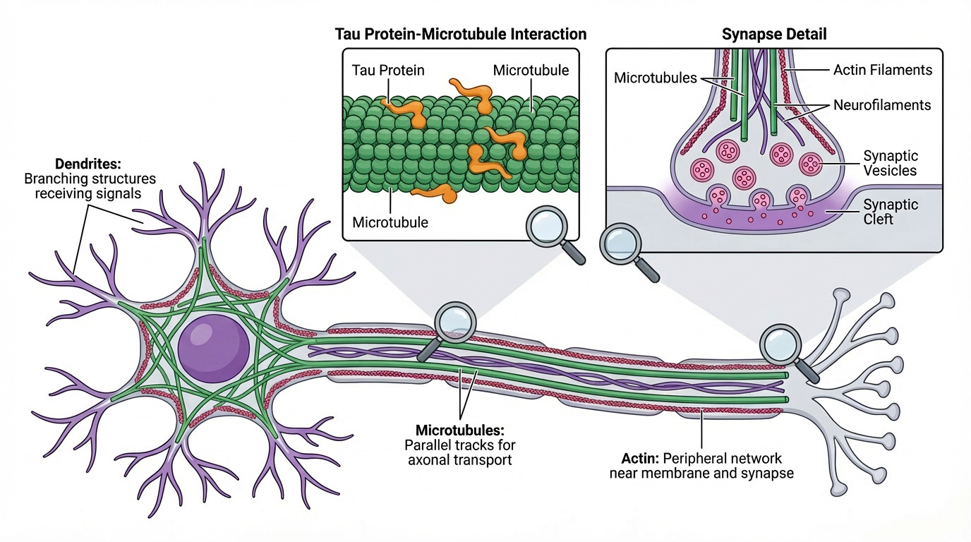
Figure 1 – Cytoskeleton of a neuron contains Microtubules (green), Actin filaments (red), and Neurofilaments (purple).
Our bodies contain many different types of living cells. Although cells are microscopic, they are highly organized and active systems. Each cell must maintain its shape, produce energy, communicate, and move materials internally in a coordinated manner. In many ways, a cell resembles a small, self-contained system with structure, transport, and constant activity.
Inside a cell, molecules and materials do not simply drift randomly. Proteins, nutrients, energy sources, and signals must travel to specific locations at specific times. This movement within the cell is known as intracellular transport. For efficient transport, the cell relies on an internal structural framework composed of specialized proteins. These structures help the cell maintain its shape and serve as tracks for intracellular transport.
Two major components of this framework are microtubules and actin filaments. Microtubules are long, tube-shaped structures that form stable tracks throughout the cell (Figure 1). Actin filaments play an important role in helping cells change shape, maintain tension, and support processes such as cell movement, cell division, and communication between neighboring cells. Together they form the cytoskeleton — a dynamic internal scaffold that supports the cell and organizes its internal activity. The cytoskeleton is present in all cells, but it is especially important in the brain, where cells must maintain complex shapes and support constant communication over long distances. Without this structural system, cells would lose their shape, and the precise movement of materials within them would be impossible.
The brain is composed of many different types of cells working together as a tightly coordinated system. Neurons are the primary signaling cells responsible for transmitting information. Alongside them are several kinds of support cells, collectively called glial cells, which protect neurons, regulate the chemical environment, provide nutrients, and maintain overall stability in the brain. These glial cells include microglia, astrocytes, and oligodendrocytes (Figure 2). Microglia serve as immune defenders, while astrocytes and oligodendrocytes help maintain neuronal health and ensure efficient signal transmission. The proper function of the brain depends on the health of all these cell types (Figure 2).
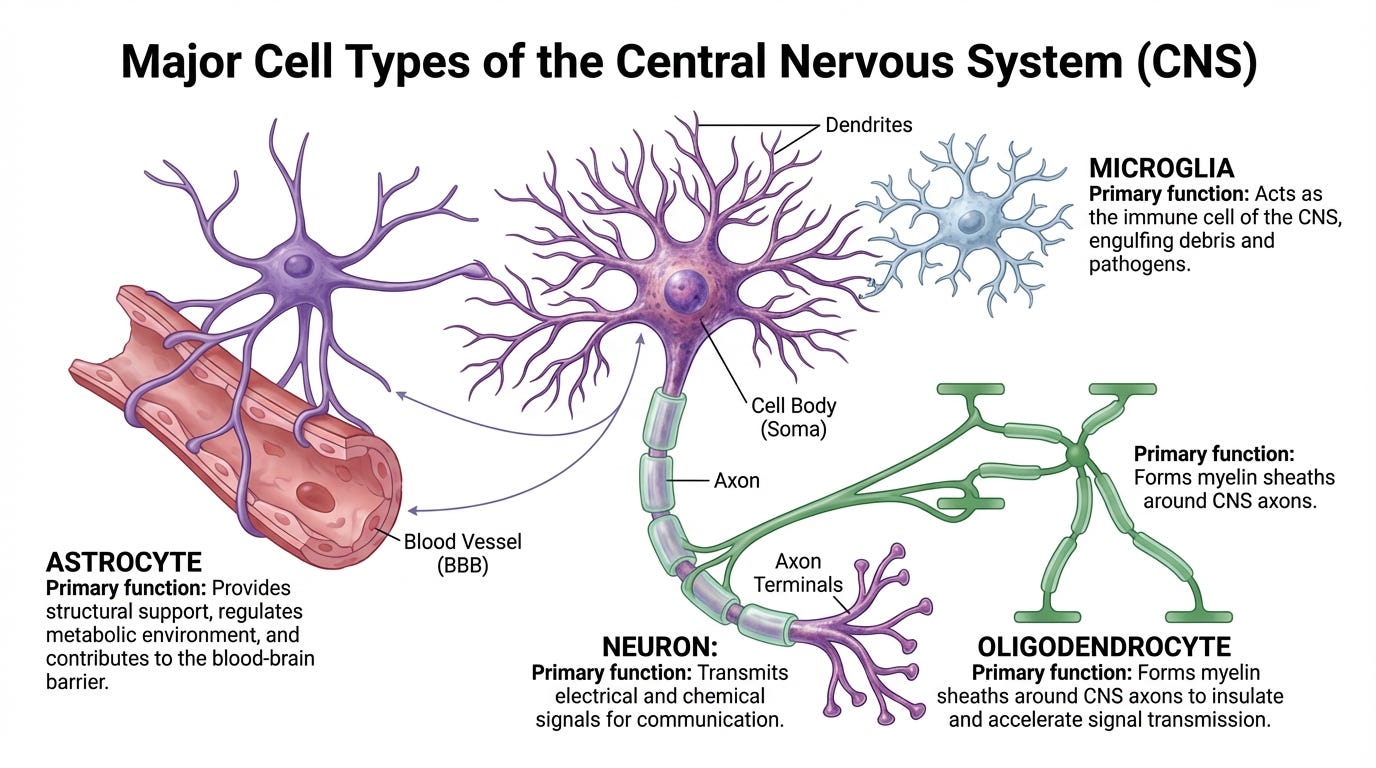
Figure 2 – Cell types in the brain showing Neurons and Glial cells (Microglia, Astrocytes, and Oligodendrocytes)
Among these, neurons are particularly specialized and uniquely vulnerable. Many neurons have extremely long extensions, allowing them to transmit signals across large distances within the brain and spinal cord. Because of this length, neurons depend heavily on efficient internal transport. Materials produced in one part of the neuron must be delivered to distant regions of the same cell, sometimes over remarkably long distances. This transport occurs primarily along microtubules, which act as stable tracks running throughout the neuron. Compared to glial cells, neurons rely heavily on long-distance transport and have limited ability to regenerate after damage, making the integrity of their intracellular transport system especially important.
When microtubules become unstable or fail to function properly, the consequences for neurons can be gradual but serious. Transport inside the cell slows or becomes disrupted. Essential materials may no longer reach critical areas, and cellular stress begins to accumulate. Over time, this can impair the neuron’s ability to function and eventually lead to cell death. Because neurons are responsible for cognition, behavior, communication, and movement, their loss directly affects brain function.
For this reason, a change affecting even a single important protein involved in maintaining neuronal structure can, over time, influence the brain’s overall health. In the following section, we will introduce one such protein — Tau — and the gene that produces it, called MAPT, and begin to explore why changes in this system can lead to disease.
In the early studies of brain biology, scientists sought to understand how neurons maintain their long shape and internal organization. During this work, researchers identified a molecule that bound strongly to microtubules, helping them remain stable. Microtubules themselves are built from repeating units of a structural protein called tubulin, which assemble into long tracks inside neurons (Figure 3). Scientists later discovered a protein that attaches along these tubulin tracks and helps regulate their stability and organization. This protein was named Tau, short for tubulin-associated unit, reflecting its close interaction with the microtubule system inside neurons (Figure 3).
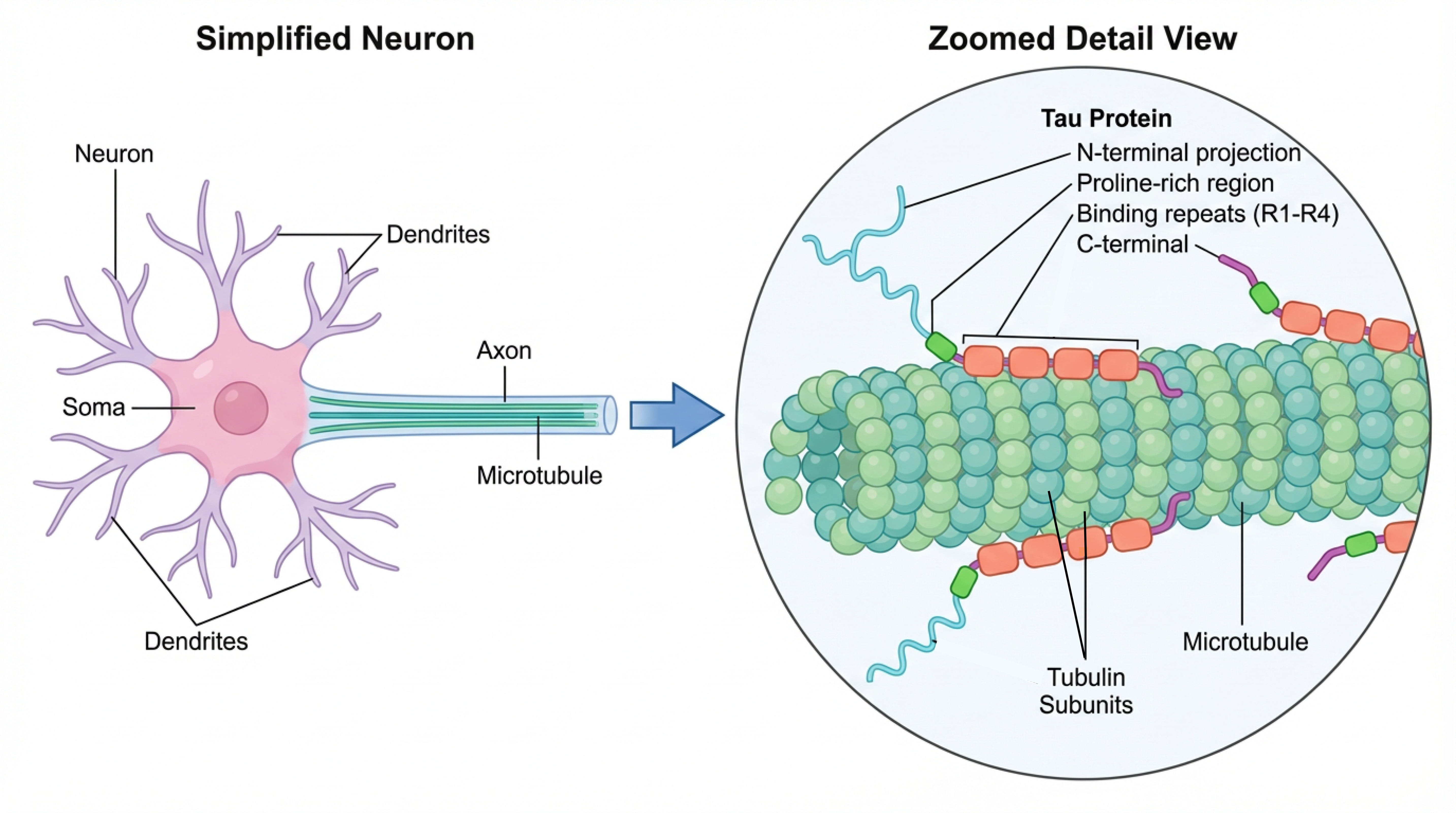
Figure 3 – Tau Protein bound to microtubules via their microtubule binding domain
Tau is produced from a gene called MAPT (Microtubule-Associated Protein Tau). To understand Tau, it is helpful to distinguish between a gene and a protein. A gene is a segment of DNA that contains instructions. A protein is the functional molecule built using those instructions. The MAPT gene provides the blueprint, and the Tau protein is the working structure that carries out its role inside the cell. Even a very small change in the gene — such as a change in a single DNA letter — can alter the structure of the protein that is produced. This change can lead to disruption in the function of the protein.
Tau’s primary role is to interact with microtubules, the long structural tracks that support intracellular transport. Microtubules must remain stable and properly organized for neurons to function over long distances and long periods of time. Tau binds along microtubules and helps maintain their stability, spacing, and organization. By doing so, Tau supports the structural integrity of the neuron and helps ensure efficient transport along microtubules. Because neurons do not readily replace themselves and must function for a lifetime, maintaining stable microtubules is especially important for their long-term survival.
To better understand how Tau performs these roles, it helps to look at how the MAPT gene is organized (Figure 4). The beginning portion of the protein, called the N-terminus, together with the short proline-rich region, helps regulate how Tau interacts with other proteins and structures within the cell. The central portion, known as the microtubule-binding domain (MBD), enables Tau to bind microtubules and directly influence their stability and organization. The ending portion, called the C-terminus, contributes to maintaining the overall structure and behavior of the protein (Figure 4). Together, these regions allow Tau to support microtubule stability, preserve neuronal structure, and help coordinate intracellular transport. Variations within these regions give rise to different forms of Tau, known as Tau isoforms, through a process called splicing.
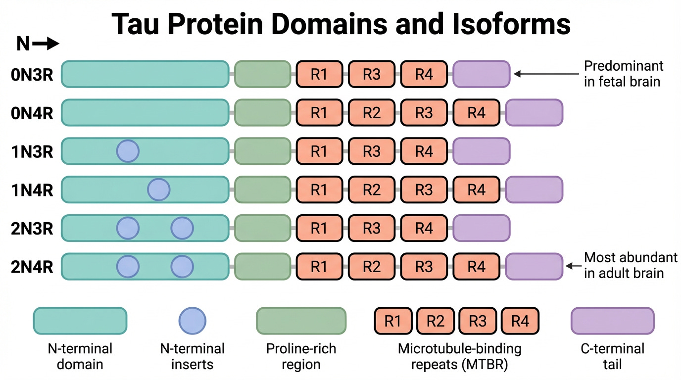
Figure 4 – Tau protein domains and isoforms
In the human brain, six main Tau isoforms are produced in healthy cells: 0N3R, 1N3R, 2N3R, 0N4R, 1N4R, and 2N4R Tau. These names reflect structural differences within the protein (Figure 4). The number before “N” refers to inserts near the beginning of the protein: 0N means no insert, 1N means one insert, and 2N means two inserts. The “R” refers to repeating segments within the microtubule-binding domain — repeated units of the region that bind microtubules. 3R contains three such repeats, and 4R contains four repeats, which influence how Tau interacts with microtubules. Variation in both the N-terminal inserts and the number of binding repeats contributes to differences in how Tau functions within neurons. Studies suggest that 4R Tau may bind microtubules more strongly and support greater stability, while 3R Tau may allow more flexibility, but the full functional differences between these forms are not yet completely understood.
The forms of Tau present in the brain change across development and aging. Before birth and during early brain development, neurons primarily produce Tau containing three microtubule-binding repeats (3R Tau). As the brain matures, neurons begin producing Tau, which contains four microtubule-binding repeats (4R Tau). In the healthy adult brain, both forms are present in a balanced state, which supports normal microtubule function (Figure 4).
Because Tau directly influences the stability and organization of microtubules, its structure is closely tied to its function. Changes that affect specific regions of the Tau protein can alter its interactions with microtubules, the stability of the cytoskeleton, and neurons' ability to maintain their structure over time. Understanding the normal structure and function of Tau provides the foundation for understanding how changes in the MAPT gene can influence neuronal health.
In the next section, we will explore how different types of changes in the MAPT gene can alter Tau and how these changes can affect its function within neurons.
No posts
Comments
Nothing yet. Say the first thing.
Sign in to join the conversation.