Jamie Andrews · The Virology Controls Studies Project
This article is going to be paid for, for 1 week, then free to read after that.
This is a very long article topping 140 pages that has taken months of research, more to this point is that whenever I have written longer articles like this with very deep research, Substack tend to Shadowban it, so having it free and open when it first goes out does me no help in just getting exposure (despite having 18k Subscribers).
I have quite a few paid for subscribers (thank you!) I am acutely aware that despite many big things going on behind the scenes there is not much you are seemingly seeing for your paid subscription, so early access can feel like it is a bit of a perk.
This will be going up as an Ebook on Shadowbanned Library for just a few dollars, if you really can’t wait you can buy the book over there. Here is a Discount code for all Substack subscribers, click there or on the image below or take an image of the QR code below that or just use code “HOAX” at the checkout.
The paid for price is set stupidly low at $35 for a years subscription, if you want to get early access then your donations are very much appreciated and are all put towards experimentally falsifying Virology.
The “Big C” has to be addressed at some point on the Virology Control Studies Project channel for the simple fact that it is the culmination of all of the hoaxes involved in Virology rolled into an endpoint Medical diagnostic of grave ill health. They have been attempting for years to tie in the fictious strands of Genetic code floating about in the air called “Viruses” with some Boogeyman “Genetic Mutation” of your cells called “Cancer”. I put “cancer” in quotation marks the same as I do “virus” at the start of anything I write to make it known that these are fraudulent terms given by the mainstream to describe the physical appearance of tissue to attribute a fake cause for disease in the whole (in some cases- apart from the asymptotic, lol).
“Cancer” is similar to a “virus” in that it does not exist as specified by the mainstream medical view of what they are. In the case of Viruses (assumed quotations from here on in) it is mislabeled cellular debris found in a cell culture from the breakdown of cell membranes and dispersion of hydrophobic membrane bound vesicles into surrounding culture medium. That breakdown can arise from imbalance at the tissue and cellular level due to the terrain of the whole being the tissue came from and/or starvation and stresses from the cell culture method. Similarly Cancer is a cellular imbalance mislabeled as a Genetic mutation that arises from a similar imbalance of the terrain of the whole. Both of these try to make the case that the disease signified (in most parts) by the cellular deformities are caused by genetics, they differ in the fact that where Viruses are a direct claimed Genetic tampering, Cancer may be “triggered” by lots of different things - Genetics, Viruses, Chemical Carcinogens, Ionizing Radiation, Heavy Metals/Metalloids and simply old age itself. At least when it comes to the formation of Cancer, for the moment they attribute it in the most part or at the very least give a sizeable amount of headroom to the notion of Terrain, environment and chemical toxicity causing Cancer. With Viruses this is pushed wholesale into the fictious story about the “arms race” of the Immune System Vs. the Foreign Invader.
Really when you lay out all of the information on a table they are the same hoax, with the same moving parts- Chemical/Environmental and Psychological stresses that cause premature disease and death blamed on a fake genetic cause that are “treated” with more Chemical stresses that cause more death and disease. Once you learn this fraudulent mechanism employed by the Allopathic death cult and their conveyor belt of death and disease you can learn how to A. Prevent and limit that initial imbalance, hence B. Not subject yourself to the Allopathic Conveyor belt C. Spot this mechanism employed in all areas of the Allopathic death cult past, present and future enabling you to protect yourself from A & B with all health decisions.
Once you fully realize the above information, you will understand that looking for a “cure” for Cancer is the wrong way of dealing with a case of imbalance. Certainly for cutting, burning and chemically poisoning ones self, which is the Allopathic way is never going to be beneficial to health. But more than that, to think you can take some sort of pill/chemical/ food stuff said to contain a chemical is also in my opinion not the way to make a change to a disease , that as we laid out above, is caused by environment, diet and lifestyle. The only way to deal with a disease caused by environment, diet and lifestyle is to… you guessed it… change your environment, diet and lifestyle. If you have been following along, the next bit should be an extension of logic in that these things, for the sake of balance, should be happening on a similar time frame as each other, a Ying to the Yang shall we put it: 3 decades of heavy drinking, smoking, processed food diets and pharma pill popping obviously cannot be undone by 3 months of cutting down on takeaways and quitting smoking whilst doing chemo (mustard gas).
So this article is a full scale falsification of the notion that Cancer/disease/ ill health can be caused by mutations of your (fake) genetics and that it can spread once you “have it” to different parts of your body like some sort of internal contagion called “metastasis”. This article started out as just focusing on the “metastasis” part in that Cancer cannot be contagious, of course, as contagion is a myth. But I think to appreciate the Metastasis Hoax we may as well include the entire Oncogenesis as a means of disease hoax in one longer format, so here we go:
As stated above, one of the main tasks set for the Virology department, certainly in the last 50 years is to culminate the Virus Hoax in a bought of deadly Cancer. The conveyor belt is easy to see: Create Fake Viral Scare> Vaccinate> Create Cancer and Fake Cancer Scare> Chemo> Die. It is a Cradle to Grave Business model of sickness for profit worth an estimated half a Trillion Dollar market cap. Not happy with this trajectory, just last year it was announced in a White House Presser by Trump and his bestest buddies Larry Ellison and Sam Altman that they were going to invest your money to the tune of another half a Trillion Dollars to build an AI Infrastructure which will “Cure Cancer”.
WASHINGTON — President Trump unveiled a $500 billion artificial intelligence infrastructure project Tuesday at the White House alongside reps from three tech and investment giants — with those business leaders asserting the initiative could cure cancer.
OpenAI, SoftBank and Oracle launched the project, called Stargate, to unleash the new technology with the help of large data centers based in Texas.
“These world-leading technology giants are announcing the formation of Stargate,” Trump said. “So put that name down in your books, because I think you’re going to hear a lot about it in the future — a new American company that will invest $500 billion at least in AI infrastructure in the United States.”
“We will see diseases get cured at an unprecedented rate. We will be amazed at how quickly we’re curing this cancer and that one — and heart disease,” added Altman, repeating that AI would “cure the diseases at a rapid, rapid rate.”
Oracle CEO Larry Ellison also expounded on the health benefits of the project.
“Little fragments of those [cancer] tumors float around in your blood. So you can do early cancer detection. If you can do it using AI, you can do early cancer detection with a blood test and using AI to look at the blood test,” Ellison said.
“Once we gene sequence that cancer tumor, you can then vaccinate the person —design a vaccine for every individual person that vaccinates them against that cancer. That mRNA vaccine, you can make that robotically, again using AI, in about 48 hours.”
He continued saying, “imagine early cancer detection, the development of a cancer vaccine for your particular cancer aimed at you and have it have that vaccine available in 48 hours. This is the promise of AI and the promise of the future.”
If we cycle back by a decade, the notion raised by Mr. Ellison that you could test for all Cancers and more from a single blood draw was by all means in motion put forward by a company called Theranos, whom unfortunately for them and CEO Elizabeth Holmes were a little too ahead of the curve:
**Theranos was a healthcare technology startup founded in 2003 by Elizabeth Holmes that claimed to revolutionize blood testing with a device called the Edison capable of running hundreds of tests from a few drops of blood. The company reached a peak valuation of $10 billion after raising approximately $700–945 million from investors, but its technology was never proven to work as advertised.
The collapse began in 2015 when Wall Street Journal reporter John Carreyrou published exposés revealing that Theranos was using traditional competitor machines for most tests and that its proprietary technology was flawed and inaccurate. This led to investigations by the FDA and CMS, which cited immediate jeopardy to patient health, causing major partners like Walgreens and Safeway to terminate their relationships with the company.
In 2018, the SEC charged Holmes and former president Ramesh “Sunny” Balwani with massive fraud, leading to the company’s dissolution. In January 2022, Holmes was convicted on four counts of wire fraud and conspiracy and sentenced to 11 years and three months in prison, while Balwani was convicted in July 2022 on ten counts of wire fraud and sentenced to 12 years and 11 months.
Unfortunately for Elizabeth if she had just waited 10 years and voted for the Orange Wotsit for a second term adjoining the first, she might not have ended up in the clink and may have just called her company Galleri (pronounced Gallery dontchaknow).
Blood tests on the horizon, and under federal regulatory view, may be about to transform cancer care — and survival.
The Food and Drug Administration has been evaluating a blood test called Galleri (pronounced like “gallery”) as a breakthrough device that can detect many different types of cancer in one test. FDA could approve it later this year, clearing the way for such testing to become broadly available.
If that happens, experts say such tools have the power to revolutionize cancer screening as it exists today — making it far easier, more comprehensive, and more accessible to a greater number of people.
Relatively few screening tests now
Currently, in the U.S. it’s common to screen for five cancers — breast, colon, cervical, prostate, and lung — each requiring its own scan, like a CT test, or exam, like a Pap smear. By comparison, the Galleri test can detect small amounts of DNA or other cancer markers for up to 50 cancers in a single vial of blood.
Galleri and another test called Cancerguard by Abbott are both multi-cancer early detection tests that already can be ordered in the U.S. under a special FDA designation. A more rigorous FDA approval process will be necessary for the tests to be covered by Medicare and other insurance.
A diagnosis will always require a follow-up exam, but the test offers solid guidance for doctors, Hall says.
“That cancer signal origin prediction is a really important feature of the technology because it directs your physician on where to go to look to confirm the presence or absence of that cancer,” she says.
Improvements in blood tests, meanwhile, dovetail with rapid developments in treatments for various cancers, making the disease ever more survivable or even curable.
Therefore, the ability to catch cancer at Stage 3 versus Stage 4 has never been more significant for survival, Hall says. “The treatment landscape has evolved so much over the past five to 10 years, that there are now a lot of treatments even for Stage 3 … and that’s a really big deal.”
Given this massive investment of your tax dollars into a previous piece of tech that landed the CEO of the bloodwork company in jail, this time it comes with Rubber Stamp approval from the State itself, so instead of pacing the yard and Tater Tots they have rolled out the Red Carpet for Celebrity Psy-op season. This in the UK mere weeks after Gelleri’s launch was Psuedo-controversial “former” BBC employer Jeremy Clarkson, who made a tear jerking announcement that he had “the Big C” at the end of his fun loving frolicking on his State subsidized farm.
He later went on to breathlessly claim on video that he was “really lucky” that he managed to “catch it early” with some bloodwork that was done, even though he “had no symptoms”…. All such a coincidence of course. He then took it up a Gear to appeal to the male bravado character that this fabulous new tech was so much better than getting a Doctor’s finger up your bum and encourage, well practically anyone with a Prostate to go and get “checked out”.
To anyone with even the slightest of discernment this was one hell of a transparent marketing push to get as many people on boarding themselves to the Allopathic death cult as possible.
Jeremy Clarkson has revealed he was diagnosed with an “aggressive” form of prostate cancer last summer and had an operation to remove 10% of his prostate, eight months after he underwent heart surgery for blocked coronary arteries.
In the final two episodes of the fifth series of his Prime Video documentary, Clarkson’s Farm, the 66-year-old presenter told his farm manager, Kaleb Cooper, and his land agent, Charlie Ireland: “I’ve got cancer.”
In the scenes, which were filmed last year and released on Tuesday night, the former Top Gear host added: “I had a medical, remember, back in May? I disappeared off the other week and I had a biopsy and it is cancer, and it’s aggressive.”
He said he “wasn’t thrilled” to receive the diagnosis but that it had been caught at a “really early stage”.
He said: “If I hadn’t have got myself checked out and they hadn’t caught the problem early, this could well have been my last harvest. It’s only because they did catch it early, there’s every hope that I’ll be harvesting this farm for many, many years to come.”
When Cooper asked him what type of cancer it was, he replied: “Where it is, is of no concern to anybody.”
But in later scenes, after undergoing a procedure, he revealed it was in his prostate. “The prostate, 10% of it’s dead,” he said. “The 10% where the cancer is.”
At the end of the series, he is filmed in a hospital bed. “So we started season five in a hospital bed and here we are at the end of season five, I’m back in a hospital bed,” he said, referring to the earlier heart operation. “Some of the treatment has gone awry, let’s say. I’ll probably be here for a little while.”
He told his viewers: “What I wanted to say was: if this is all successful, I’ll see you for season six. And if it isn’t, I won’t. Take care, everyone.”
On Tuesday evening he warned fans on social media he had some “sombre news” with regard to Clarkson’s Farm. “Ordinarily we try to keep the show bucolic and charming and cheerful. But the final two episodes which drop in the middle of the night tonight are none of those things, really – they’re a difficult watch.”
He sounded emotional as he added: “They’re really, really difficult.”
Following the episodes’ release, charity Prostate Cancer UK thanked Clarkson for sharing his diagnosis and treatment saying it would raise “vital awareness”.
Chiara De Biase, the charity’s fundraising and health strategy director, said: “Thankfully he found the disease at an early stage, but sadly this is still not the experience of many men across the UK.
“Over 10,000 dads, brothers, sons and friends are diagnosed too late for a cure every year, and today the responsibility to know if you’re at higher risk and to act on it rest entirely on men’s shoulders. This must change.
“Any men worried by Jeremy’s story should take our 30 second online risk checker or have a chat with their GP about a quick and simple blood test to check for any signs.”
Prostate cancer is one of the most common cancers in the UK, accounting for 28% of all new male cancer cases, according to Cancer Research UK.
Last June, Clarkson had urged men to go for prostate checkups, saying: “I’ve had too many friends go down with prostate cancer, and all it takes to get on top of the situation early is a moment or two of being a bit cross-eyed. You get the all-clear and the doc goes home happy. What’s not to like?”
There is no national NHS screening programme for prostate cancer, but men aged 50 or over – and those aged 45 or older who have a strong family history of prostate cancer or are of a Black or African-Caribbean ethnicity – can request a prostate-specific antigen (PSA) blood test from their GP.
We can see the exact trajectory of this thing, which is the identical roadmap of 2020, mass screening leads to Cancer Pandemic. This is obviously not going to be quick peaks as with this hoax they are going to slowly ramp this mother up until they fulfil their quote of everything with a pulse has Cancer. They have not deviated one iota away from the tools used in the Scamdemic, in that they are using PCR and Genomic sequencing as the front of house triage. As explained on this channel in depth, the PCR test is a group of reagents that act like a blank canvas, you can effectively achieve whatever result you wish in assay design by altering the intrinsic threshold that the fluorescent lights will go ping.
So we can update the flow chart as such: Create Fake Viral Scare>Fake PCR and Genetics test> Vaccinate> Create Cancer and Fake Cancer Scare>Fake PCR and Genetics test> Chemo> Die. With this model it is a bit like Quantitive Easing where Central Banks just print digital money and “inject” it into economy. Why not just put 6 zeros on the end of the number you just invented, it literally makes no difference, right?
The scientist most often credited with the original idea is Theodor Boveri.
In 1902–1914, Boveri studied sea urchin embryos under a microscope. He experimentally created embryos with abnormal numbers of chromosomes and observed that they developed abnormally. From this, he reasoned that chromosomes carry hereditary information and that cancers might arise when a cell acquires the wrong combination of chromosomes, causing uncontrolled growth.
What’s remarkable is that Boveri reached this conclusion before DNA was known to be the genetic material and decades before scientists could directly identify mutations. His proposal was largely theoretical, based on chromosome behavior and careful observation.
How the idea was confirmed
Boveri’s hypothesis wasn’t fully accepted until much later.
1. Chromosome abnormalities were found in cancer cells
During the mid-20th century, improved microscopy and cytogenetics revealed that many cancers consistently contain abnormal chromosomes. Scientists found specific chromosome changes associated with particular cancers.
One famous example was the discovery by Janet Rowley that a characteristic chromosome translocation occurs in chronic myelogenous leukemia, providing strong evidence that genetic alterations can drive cancer.
Boveri’s methods were ingenious for the time, because he had no molecular biology tools, no DNA sequencing, and no way to manipulate genes directly. He relied on experimental embryology and microscopy.
The key experiment: creating embryos with abnormal chromosome numbers
Boveri worked with the eggs of the sea urchin Sea urchin.
Normally:
One sperm fertilizes one egg.
The resulting embryo gets a normal set of chromosomes.
Development proceeds normally.
Boveri experimentally induced polyspermy—fertilization of a single egg by two sperm instead of one.
How he did it
He altered seawater conditions around freshly spawned sea-urchin eggs, increasing the frequency with which multiple sperm entered the same egg. (The exact procedures varied among experiments, but generally involved manipulating fertilization conditions rather than surgically altering chromosomes.)
The result was:
Eggs receiving two sperm had three centrosomes instead of the normal two.
During cell division, this often produced tripolar or multipolar spindles rather than the normal bipolar spindle.
Chromosomes were distributed unevenly among daughter cells.
Under the microscope, Boveri could directly observe these abnormal divisions.
What he observed
Instead of every daughter cell receiving a complete chromosome set, some cells received:
Too many chromosomes
Too few chromosomes
Different combinations of chromosomes
The embryos often developed abnormally or died.
This was important because many scientists at the time thought chromosomes were largely interchangeable. Boveri’s results suggested that individual chromosomes have distinct functions, because losing particular chromosomes caused specific developmental failures.
The logical leap to cancer
Boveri reasoned:
Abnormal chromosome combinations can produce cells with abnormal behavior.
A multipolar division occurring in a normal tissue could create a cell with a dangerous chromosome combination.
That cell might then proliferate uncontrollably.
Therefore, tumors could originate from chromosome abnormalities within a single cell lineage.
Today we’d describe this as a hypothesis that cancer arises from somatic genetic alterations.
An important caveat
Boveri did not directly create cancer in sea urchins.
His evidence was indirect:
He demonstrated that chromosome abnormalities can fundamentally alter cell behavior and development.
He observed chromosome abnormalities in many tumor cells reported by pathologists.
He proposed that the same principle could explain malignant growth.
The direct demonstration that specific genetic changes cause cancer came much later, through:
Cancer cytogenetics (e.g., specific chromosomal translocations in leukemia),
Viral oncogene studies,
Discovery of oncogenes and tumor suppressor genes.
So Boveri’s experiments were less “I induced cancer” and more “I showed that changing chromosome composition profoundly changes cell fate, and therefore cancer could be a chromosome disease.”
Q: A sperm triggers a rise in calcium in the ovum when it enters, correct? If two sperm enter one egg would there be chaotic calcium signaling and how might that affect the eggs development?
Yes—when a sperm enters the egg, it typically triggers a rise in intracellular calcium, usually as a series of oscillations rather than a single spike. In mammals this is driven mainly by the sperm-delivered factor PLCζ, which causes repeated release of calcium from the egg’s internal stores. These calcium oscillations are what “activate” the egg and start embryonic development.
If two sperm enter the same egg (a condition called polyspermy), the situation doesn’t usually produce a clean “double-strength” calcium signal. Instead, the normal regulatory sequence is disrupted.
Here’s what tends to happen:
Each sperm can contribute activating signals, so you may get abnormally strong or irregular calcium oscillations.
The egg is designed to respond to a tightly timed pattern of calcium spikes; multiple inputs can make the pattern less coordinated or prolonged.
More importantly, the biggest problem is not calcium itself but chromosome balance: the egg now has extra sets of paternal chromosomes, which leads to severe genomic imbalance.
Even if calcium activation occurs, polyspermic embryos almost always fail because:
The spindle cannot properly organize multiple haploid sets.
Cell divisions become abnormal.
Development arrests very early.
To prevent this, eggs have strong anti-polyspermy defenses:
A fast block (brief membrane depolarization in some species)
A slow block, where calcium-triggered cortical granules modify the egg’s outer layer to prevent further sperm entry
So while multiple sperm could in principle disturb calcium signaling, the dominant reason polyspermy is lethal is that it produces an unworkable chromosome set, not primarily because of calcium chaos.
Boveri more than half a century before they invented DNA, started to observe real, physical things, i.e Chromosomes. As explained in many other writings, Chromosomes are just physical structures that are the result of the separating of an Ionic solution. No much more than for instance seeing strings of melted cheese when you pull a hot Jalapeno Popper apart. The movement creates patterns of shear and elastic stresses in the gel phase in between solid and liquid. The different patterns (Chromosomes) are made if the consistency of the phase changes through altering the Ionic Concentration inside the membrane of the cell.
This was clearly acutely apparent to Boveri whom when trying his Sea Urchin Swinger’s Party made sure to use “Calcium Free Sea Water”. As with most of these “sciences” they start out making mostly real observations in completely inconsequential things and draw these strange all encompassing conclusions that are then double/tripled down on in the future, seemingly picked at random by people with Tests.
So what did actually learn from Boveri’s work? Well that if you take normal cells that you can observe reproducing and force more in that they do in nature, then the resultant cell is deformed. Why anyone felt the need to expand on this basic piece of logic is a bit baffling, considering that they already knew full well that altering the charge inside the Ovum but inducing Calcium release, if done to extremes arrests the development of the Ovum. They already had the mechanism of both reproduction and destruction; charge.
Alfred G. Knudson proposed in 1971 that cancers like retinoblastoma require two genetic “hits” (mutations) before a normal cell becomes cancerous.
This became known as the two-hit hypothesis.
📄 His key paper
Original paper (1971): “Mutation and cancer: statistical study of retinoblastoma”
This paper is one of the most famous in cancer genetics.
🔬 What Knudson actually did (methods)
Importantly, Knudson did not use molecular lab experiments. His work was based on:
1. Epidemiological analysis
He studied cases of:
Retinoblastoma
He compared:
Early-onset cases (often bilateral tumors)
Later-onset cases (usually unilateral tumors)
2. Statistical modeling
He analyzed:
Age of diagnosis distributions
Frequency of single vs. multiple tumors
Population incidence rates
He then asked:
Could the timing and pattern of disease be explained by mutation accumulation?
3. Mathematical inference of mutation steps
He proposed two distinct mechanisms:
(A) Hereditary form
Child inherits one defective allele (first “hit” already present)
Only one additional somatic mutation is needed
→ earlier onset, often both eyes affected
(B) Sporadic form
Both mutations must occur in the same retinal cell lineage
→ later onset, usually one eye
4. Logical reconstruction of mutation timing
He modeled tumor formation as a multi-step stochastic process, where:
Each “hit” is a rare mutation event
Cancer risk increases with time as mutations accumulate
This was one of the first quantitative frameworks linking:
cancer = accumulation of genetic mutations in somatic cells
🧬 Why this was revolutionary
Before Knudson:
Cancer was often seen as “one catastrophic event”
After Knudson:
Cancer became understood as a stepwise genetic process
His model directly supported later discoveries of:
tumor suppressor genes (like RB1)
somatic mutation accumulation
multi-hit carcinogenesis
ABSTRACT Based upon observations on 48 cases of retinoblastoma and published reports, the hypothesis is developed that retinoblastoma is a cancer caused by two mutational events. In the dominantly inherited form, one mutation is inherited via the germinal cells and the second occurs in somatic cells. In the nonhereditary form, both mutations occur in somatic cells. The second mutation produces an average of three retinoblastomas per individual inheriting the first mutation. Using Poisson statistics, one can calculate that this number (three) can explain the occasional gene carrier who gets no tumor, those who develop only unilateral tumors, and those who develop bilateral tumors, as well as explaining instances of multiple tumors in one eye. This value for the mean number of tumors occurring in genetic carriers may be used to estimate the mutation rate for each mutation. The germinal and somatic rates for the first, and the somatic rate for the second, mutation, are approximately equal. The germinal mutation may arise in some instances from a delayed mutation.
PATIENT DATA The records of all retinoblastoma patients admitted to the M. D. Anderson Hospital, some 48 cases during the period 1944-1969, were reviewed. These cases are tabulated (Table 1) with respect to unilaterality or bilaterality, sex, age at diagnosis, and family history. Whenever possible, the number of tumors in each eye was estimated.
Ahem (clears throat), so this is one of the seminal papers showing that Cancer is caused by checks notes Genetics. Really we need look no further than the title being that it is a Statistical analysis, therefore is under the umbrella of Epidemiology and is hence absolutely and totally worthless in assessing causality. Epidemiology assumes causation and works off of this as a spring board, it is the literal definition of correlation ≠ causation.
It’s kind of alot worse than this given the absolute torture of the statistics. They have started with a cohort from a local hospital of just 48 kids spanning over the course of a quarter of a century. They take all these kids which have blemishes on their eyes, tell these toddlers they have cancer and then posthumously number crunch them to fit a predefined assumption. You have to assume that all records of family members are absolutely correct over this very long period and that everyone of the family members has been thoroughly clinically examined, neither of which should be assumed to be accurate as Knudson has compounded in his errors.
Anything that uses “Poisson Statistics” (French for- Fish, of which they attribute to pranking given that April Fools Day is Poisson D’Avril) to find a “rule of thumb” should be openly mocked as complete quackery.
J. Michael Bishop and Harold Varmus are best known for showing that cancer-causing genes (“oncogenes”) are actually normal cellular genes that have been altered, not foreign viral genes. This was a major shift in cancer biology.
🧠 What they discovered (core idea)
Before their work:
Scientists thought oncogenes were only carried by cancer-causing viruses
After their work:
Oncogenes are normal human genes (“proto-oncogenes”) that can become cancer-causing when mutated or misregulated
This helped establish cancer as a genetic disease of the cell itself, not something always introduced from outside.
📄 Key papers
1. The famous SRC gene discovery (1976–1978 work leading to model)
A foundational paper:
Stehelin et al., 1976 (Cell) “DNA related to the transforming gene(s) of avian sarcoma viruses is present in normal avian DNA”
viral oncogenes are modified versions of normal genes
5. Comparative evolutionary reasoning
They also used logic across species:
If normal animals have homologous genes to viral oncogenes
then those genes must be ancient cellular genes
This supported the “proto-oncogene” concept.
🧬 What they proved conceptually
They established three key ideas:
1. Proto-oncogenes exist in normal cells
Normal genes like src, ras, myc
2. Cancer arises when these genes are altered
mutation
overexpression
viral capture
3. Viruses “steal” cellular genes
Retroviruses can incorporate host genes and modify them
🏆 Impact
Their work helped lead to:
the concept of oncogenes
the idea that cancer is fundamentally a genetic disease of somatic cells
the 1989 Nobel Prize in Physiology or Medicine (Bishop & Varmus shared it)
DNA related to the transforming gene(s) of avian sarcoma viruses is present in normal avian DNA Infection of fibroblasts by avian sarcoma virus (ASV) leads to neoplastic transformation of the host cell. Genetic analyses have implicated specific viral genes in the transforming process¹⁻⁴, and recent results suggest that a single viral gene is responsible⁴. Normal chicken cells contain DNA homologous to part of the ASV genome⁵⁻⁸; moreover, embryonic fibroblasts from certain strains of chickens can produce low titres of infectious type C viruses either spontaneously⁹ or in response to various inducing agents¹⁰. None of the viruses obtained from normal chicken cells, however, can transform fibroblasts, and results with molecular hybridisation indicate that the nucleotide sequences responsible for transformation by ASV are not part of the genetic complement of the normal cell¹¹. We demonstrate here that the DNA of normal chicken cells.
“Here have a Nobel Prize for failing a Control and making some shit up”
We really don’t need to delve very far into this cacophony before we see them unload a 12 gauge shotgun straight into their toes. This more modern paper that came out ever so slightly before the invention of Sanger Sequencing piled even more assumptions onto the Everest amount of assumptions from their predecessors. How they thought that staining a gel with some dye and battery electrodes could infer anything is a constant source amusement especially given the absolute clanger that they introduce the paper with, i.e the fact that they find the same genes they are looking for in healthy animals, i.e they already failed their control.
Of course any intellectually honest people would have stopped there, packed up their bags and gone home, but not this lot. For a start it was pretty hilarious, but still unsurprising, that they were studying the “genetics” of fictitious “cancer” causing “viruses” in the first place. Add to this that they found this claimed “sarcoma Virus” in healthy birds, but strangely no mammals and you have the full picture for the crazy fairytale of Oncology and Virology. They invented the idea that this Cancer causing Virus was just standard part of a birds genetics and this means somehow that therefore genetics cause Cancer. It is such an art to be able to turn abject failure into somehow into a win, but Scientists especially Nobel Prize winning artists are masters at it seemingly.
A fundamental challenge to SMT lies in its quantitative feasibility. Using a Poisson-Erlang waiting-time model, Olsen and Liisberg (2025) managed to out Fish their predecessors Fish when they tested whether empirically observed mutation rates and cell-division frequencies permit multi-hit carcinogenesis within biologically realistic timescales
Their analysis reveals a striking conclusion: even under optimistic assumptions, expected waiting times exceed progenitor-cell lifespans by several orders of magnitude.
The calculation is instructive. Each cell division introduces random copying errors at approximately 10⁻⁹–10⁻⁸ per base pair. Only a minute fraction of the genome—roughly 10³–10⁵ base pairs—can yield cancer-relevant driver effects when altered. SMT assumes that a single somatic cell lineage must sequentially accumulate five to seven independent driver mutations within a human lifetime
.
The limiting factor is lineage lifespan, not mutation frequency. Progenitor cells divide rapidly but exist too briefly (approximately six divisions before differentiation). Stem cells persist but divide infrequently (roughly 70 times per year). No lineage provides the temporal window required for stochastic multi-hit accumulation. This quantitative analysis renders sequential multi-hit carcinogenesis “statistically and biologically impossible.
Summary
The mutation paradox exposes a deep logical tension at the core of the Somatic Mutation Theory.
The very cells most consistent with SMT’s probabilistic requirements - stem and progenitor cells - already possess intrinsic proliferative ability and depend on contextual signals for regulation. Mutations cannot “add” proliferation to these cells; they can only erode the communication that normally restrains it, a process far more complex than SMT’s linear, gene-centric model allows. Empirical evidence that environmental normalization can restore malignant cells to order underscores the inadequacy of cell-autonomous explanations.
The next section broadens this analysis by situating carcinogenesis within the organism’s hierarchical organization, showing how proliferation, differentiation, and repair emerge from multi-scale communication rather than from isolated genetic events.
The Statistical and Conceptual Limits of the Somatic Mutation Theory
The Somatic Mutation Theory (SMT) provides a compelling narrative: a single somatic cell accumulates a sequence of random mutations that, over time, confer the hallmarks of malignancy.
Yet when the theory is evaluated against both quantitative constraints and biological organization, its explanatory power sharply declines. The preceding sections demonstrated that the probability of acquiring the necessary combination of driver mutations within a single lineage is vanishingly small, and that the cells most compatible with SMT’s requirements - stem and progenitor cells - already possess regulated proliferative capacity. Together, these observations reveal that SMT is limited not only by statistics, but also by logic: the model presumes both an improbable sequence of events and an implausible cellular context.
This mismatch between probability, lineage identity, and observed phenotype delineates the limits of a cell-autonomous view and motivates the shift toward a systems-regulatory framework.
The statistical boundary
The Poisson/Gamma waiting-time model clarifies the temporal horizon within which mutation-driven carcinogenesis could occur. Even under favorable assumptions - a mutation rate of 10-8 per base pair per division, a large mutational target (105 bp), and numerous parallel lineages - the expected waiting time for five to seven driver mutations remains on the order of months to years. Under realistic physiological conditions, it extends to centuries or millennia.
A pretty big whoopsie has occurred in modern times when looking at the Somatic Mutation Theory and using all of the Science Establishments own Fishy mathematics. In that the rate it would take for Cancer mutations to occur would be less like months and years and more like Centuries and Millenia. It’s a funny thing that happens when you go with their mountain of assumptions and just plug their own numbers into their own equations and find out that they were out by factors of 100, it’s as if they pulled these theories out of their backsides in the first place.
The “Unnamed Cell” problem is a major problem of the Somatic Mutation Theory (SMT) of cancer, highlighting its vagueness from the fact that it was largely a figment of someone’s active imagination. Yes, control experiments have been conducted that challenge SMT’s predictions and would you have it, they fail, repeatedly.
The Somatic Mutation Theory (SMT) posits that cancer is a cell-autonomous disease, initiated by an accumulation of random mutations in a single cell’s DNA that cause it to proliferate uncontrollably. The “Unnamed Cell” points out that while SMT talks about a “normal cell” transforming, it fails to specify which kind of normal cell. Most cells in the human body, like neurons and muscle fibers, are terminally differentiated and cannot divide. Therefore, the cell that accumulates mutations must be one that retains the ability to proliferate—specifically, adult stem cells or progenitor cells . The problem is that SMT rarely makes this crucial distinction explicit. By leaving the “initiating cell” unnamed, the theory avoids the biological constraints that specifying a target cell would impose, such as the tissue’s specific control mechanisms. This vagueness allows SMT to appear universally applicable while sidestepping a key question: if a cell is already capable of regulated proliferation, what specific change makes it cancerous? It’s a reoccurring theme within Soyence that of a complete lack of Specificity.
The “fitness paradox” in cancer biology describes a counterintuitive reality: mutations that we typically label as “cancer-causing” do not inherently make cells stronger or “fitter.” In fact, within a healthy tissue environment, these mutations often make cells less fit than their normal neighbors. A mutation’s value is not absolute; it is entirely dependent on the surrounding cellular ecosystem. This challenges the simple genetic view of cancer and is best understood through three interconnected principles.
1. Fitness is Context-Dependent, Not Inherent
The central idea is that the fitness of a cell is a property of its relationship with the environment. Normal cells have undergone millions of years of evolution to be exquisitely adapted to the healthy tissue environment. In this “lawn” of healthy cells, any random mutation is statistically more likely to disrupt this fine-tuning and reduce fitness
.
A Key Example: Take the PIK3CA mutation, a common oncogenic driver. In a healthy esophagus, a cell with this mutation can gain a slight competitive advantage and expand. However, this advantage is not fixed. Research has shown that the expansion of PIK3CA mutant clones is heavily influenced by the body’s metabolic environment.
In conditions like type 1 diabetes or diet-induced obesity, the mutant cells’ competitive fitness is enhanced, allowing them to expand more rapidly.
Conversely, the anti-diabetic drug metformin was able to reduce this mutant advantage. This clearly demonstrates that the “fitness” of an oncogenic mutation is malleable and responds to systemic changes like diet.
2. The “Decoy Peak” and Aging Tissue
This context-dependency explains why many cancer-associated mutations are found in healthy tissues without causing cancer. Evolutionary biologist James DeGregori and others have proposed an “Adaptive Oncogenesis” model to explain this.
The Model: Healthy, young tissues are like a high, stable “fitness peak” that normal cells are optimized to occupy. Any mutation pushes a cell down this peak, decreasing its fitness
Aging Changes the Landscape: As we age, the tissue environment degrades due to accumulated damage and changes in the microenvironment. This lowers the “fitness peak” for normal cells, creating a new landscape where certain mutations can now confer a relative advantage.
The Decoy Peak: Some mutations, like those in NOTCH1, can place a cell on a lower, safer “decoy peak”
. These cells are more competitive in an aging tissue but have a low risk of progressing to cancer. They may even actively suppress tumor growth, which is why NOTCH1 mutations are common in healthy aging esophagus but rare in esophageal cancers. This suggests that clonal expansions in aging tissues are often a normal part of tissue dynamics and not, by themselves, a sign of impending cancer
.
3. Implications: A Shift from “Targeting Mutants” to “Supporting Health”
The fitness paradox has profound implications for how we think about cancer treatment and prevention. The current focus is on developing drugs that directly kill cells with specific mutations. However, if a mutation is only dangerous in a damaged landscape, a more effective strategy might be to “fertilize the lawn” —improving the health of the tissue environment to restore the fitness advantage of normal cells
.
This model elegantly explains Peto’s Paradox: If cancer were purely about random mutation accumulation, large, long-lived animals like whales should have astronomically higher cancer rates. They do not, because their tissues have evolved robust mechanisms to maintain a healthy landscape that suppresses the emergence of advantageous mutants.
The fitness paradox tells us that a mutation is only as dangerous as the environment allows it to be. This reframes cancer not as a genetic inevitability, but as a disease driven by the systemic and environmental context in which cells live.
Q: Are there any documented examples of wild Galapagos tortoises having cancer?
No. There are currently no clearly documented, published cases of a free-ranging wild Galápagos tortoise being diagnosed with cancer while living in the wild.
There are documented cases of cancer in Galápagos tortoises, including a mast cell tumor reported in a tortoise that had originally been captured in the wild but was living in a breeding center when the tumor was discovered and diagnosed.
So the concise answer is:
No documented published example of a wild, free-ranging Galápagos tortoise with cancer; documented cases are from tortoises in captivity or managed breeding programs.
It’s wonderful when not only you are comparing made up parameters with made up parameters within one small section of a major branch of science, but when you start to compare these things holistically is when the deep chasms of terrible logic reveal themselves. If these are in general “random mutations” then all old things are riddled with cancer. You’d basically only see a large tumor in a Tortoise Shell walking around the Galapagos Islands.
Funnily enough you never see “mutations” that are positive, they are always defects and by this very notion we can safely chuck the notion of “random mutations” in the bin, no matter what is the driving mechanism behind them. There is adequate natural growth and their is injury and deformity it’s as simple as that. So a two-fer in this tiny little section debunking Evolution and Genetic Mutations in one with some simple logic.
This refers to a series of experiments where malignant cells were placed in a normal tissue microenvironment, causing them to revert to a normal, non-cancerous phenotype
. This demonstrates that cancer is not an irreversible cell-autonomous state, as SMT would suggest, but is heavily influenced by signals from the surrounding tissue.
CANCEROUS cells can be forced to behave like normal cells by fine-tuning their chemical communication with their environment, say researchers in the US. They hope that this startling discovery could add a new dimension to breast cancer research.
Mina Bissell and Valerie Weaver of the Lawrence Berkeley National Laboratory in California and their colleagues made the discovery after looking at the way breast cells group together. In normal breast tissue, around 8 cells cluster into spherical groups, each surrounded by a fibrous protein mesh. But in cancerous breast tissue, in which the cells divide uncontrollably, this mesh is irregular and the cells clump together randomly.
The team made three-dimensional cultures of breast cells. They then added an antibody to a receptor called beta-1 integrin, which allows cells to “communicate” chemically with the extracellular matrix, the gel of proteins between groups of cells. They found that the antibody linked up with tumour cells in a more haphazard pattern than with normal cells, suggesting that the beta-1 integrin molecules are not in the right places on the cell surfaces. The tumour cells also had more receptors than usual.
Advertisement
The researchers report in the current issue of the Journal of Cell Biology that tumour cells soaked in an antibody that blocks beta-1 integrin undergo a dramatic change, and become normal in appearance and behavior. “At first I thought I had mixed the normal and tumour cell tubes up,” says Weaver.
Repeats of the experiments confirmed that although the reverted cells still harbour the genetic mutations that made them cancerous, they stop dividing uncontrollably and regroup in normal clusters. Weaver suggests that this return to the normal structure stops the cells dividing like tumours.
Until now, cancer research has been dominated by the study of genes that trigger cancer when switched on, and those that protect against cancer. Ray White of the University of Utah’s Huntsman Cancer Institute in Salt Lake City foresees a change, however. “Bissell’s research opens up a third area of regulation of cells through their interactions with their outside environment,” he says.
Abstract.
In a recently developed human breast cancer model, treatment of tumor cells in a 3-dimensional culture with inhibitory b1-integrin antibody or its Fab fragments led to a striking morphological and functional reversion to a normal phenotype. A stimulatory b1-integrin antibody proved to be ineffective. The newly formed reverted acini re-assembled a basement membrane and re-established E-cadherin–catenin complexes, and re-organized their cytoskeletons. At the same time they downregulated cyclin D1, upregulated p21cip,waf-1, and stopped growing. Tumor cells treated with the same antibody and injected into nude mice had significantly reduced number and size of tumors in nude mice. The tissue distribution of other integrins was also normalized, suggesting the existence of intimate interactions between the different integrin pathways as well as adherens junctions. On the other hand, nonmalignant cells when treated with either a6 or b4 function altering antibodies continued to grow, and had disorganized colony morphologies resembling the untreated tumor colonies. This shows a significant role of the a6/b4 heterodimer in directing polarity and tissue structure. The observed phenotypes were reversible when the cells were disassociated and the antibodies removed. Our results illustrate that the extracellular matrix and its receptors dictate the phenotype of mammary epithelial cells, and thus in this model system the tissue phenotype is dominant over the cellular genotype.
There are again a couple of points to make with this one paper. Whatever they are using as an “antibody” titer is clearly supplemented with growth medium or somehow provides sustenance to the cell line as this has all of the hallmarks of the cell culture isolation fraud with happy/sad cells just based on their dietary modifications. It is hard to really pinpoint this as, unsurprisingly, there is no method for the making of the “Antibodies”. Whichever way you skin it they are adding something demonstrably non genetic that seems to undo what they claim is a disease caused by genetics. That’s a big problem, of course the story will be that it does genetic magic to undo the genetic terror, but they can make up whatever stories they like.
The compounding problem here is the fact that genetic mutations are meant to be permanent, yet here we have the proverbial Petri Dish Quasimodo that if you rub a little of the special tincture into his face, he is now Brad Pitt. This is pretty compelling evidence that Cancer (cell deformation) is just a state of balance. I will describe what is in my mind thusly. Take a balloon that represents the cell membrane. If you fill it with water, it will fill the balloon normally. If you filled it with Jam (strawberry preserve) it would sag and warp as the gel like consistency from the pectin clumps rather than provides equal hydrostatic pressure. If you put Mercury in the balloon it would burst under its own weight.
The absolute essence of Scientific falsification as we have become accustomed to by now is the humble but almighty Control. I wouldn’t have called my channel what it is, if I didn’t believe in what this simple logic gate does for revealing the truth about a subject. So really if we want to learn about the incidence of claimed “genetic mutation caused Cancer” we need to look at healthy tissue from healthy people and sequence it for genetic mutations. The key, paradigm-shifting finding from multiple control studies is that by middle to old age, a staggering proportion of cells in normal, cancer-free tissues harbor what would traditionally be called “cancer-driving” mutations.
Meta-Analysis of Normal Tissues: A landmark meta-analysis combined data from sequencing studies across multiple healthy tissues—including colon, endometrium, esophagus, skin, and blood . It revealed that in older age groups, over 50% of cells in these tissues had mutations in known cancer genes . The study calculated that an average older, cancer-free individual possesses at least 100 billion cells that carry at least one oncogenic mutation . This finding completely overturns the idea that these mutations are rare, early, and specific warning signs of cancer.
Abstract
BACKGROUND
To shed light on the earliest events in oncogenesis, there is growing interest in understanding the mutational landscapes of normal tissues across ages. In the last decade, next-generation sequencing of human tissues has revealed a surprising abundance of cells with what would be considered oncogenic mutations.
AIMS
We performed meta-analysis on previously published sequencing data on normal tissues to categorize mutations based on their presence in cancer and showcase the quantity of cells with cancer-associated mutations in cancer-free individuals.
METHODS AND RESULTS
We analyzed sequencing data from these studies of normal tissues to determine the prevalence of cells with mutations in three different categories across multiple age groups: 1) mutations in genes designated as drivers, 2) mutations that are in the Cancer Gene Census (CGC), and 3) mutations in the CGC that are considered pathogenic. As we age, the percentage of cells in all three levels increase significantly, reaching over 50% of cells having oncogenic mutations for multiple tissues in the older age groups. The clear enrichment for these mutations, particularly at older ages, likely indicates strong selection for the resulting phenotypes. Combined with an estimation of the number of cells in tissues, we calculate that most older, cancer-free individuals possess at least a 100 billion cells that harbor at least one oncogenic mutation, presumably emanating from a fitness advantage conferred by these mutations that promotes clonal expansion.
CONCLUSIONS
These studies of normal tissues have highlighted the specific drivers of clonal expansion and how frequently they appear in us. Their high prevalence throughout cancer-free individuals necessitates reconsideration of the oncogenicity of these mutations, which could shape methods of detection, prevention and treatment of cancer, as well as of the potential impact of these mutations on tissue function and our health.
When investigating oncogenesis throughout the human body, it is important to consider a life history perspective – how natural selection has shaped our tissues to maximize survival and reproductive success and the limits of these selective pressures at older ages when we are less likely to reproduce.12 In this light, we can better appreciate why some clonal expansions may be more tolerated than others and the age-dependence of this tolerance. Due in large part to the fact that our lifespan has increased significantly in recent centuries, age has become the biggest cancer risk factor and is a crucial component to investigating the transition from clonal evolution to oncogenesis.1,13–15 Attention must also be paid to the fact that different turnover rates and mutation rates exist across epithelial tissues and the hematopoietic system, which when combined with very different microenvironment-driven selective pressures, leads to differing clonal evolution routes to cancer. Therefore, providing insight on the somatic mutational landscape and its potential to progress to cancer throughout the human body across age is critically important.
Stroma-Dependent Carcinogenesis: In one study, normal mammary epithelial cells were transplanted next to stroma (connective tissue) that had been previously exposed to a carcinogen. These normal cells gave rise to tumors at a much higher rate than controls. This shows that the tissue environment (the stroma), not just mutations within the epithelial cells themselves, can be a primary driver of cancer.
Abstract
Understanding the early stages of carcinogenesis requires detailed insight into the abnormalities present in normal cells before cancer onset. In the past, it was difficult to analyze genomic abnormalities in small clones in normal tissues. However, recent technological advances in genomic analysis have shed light on the process of accumulation of somatic mutations in normal cells, which is driven by factors such as aging and environmental influences. Even in normal tissues, clones that have acquired driver mutations—either directly contributing to carcinogenesis or adapting to specific pathological or genetic backgrounds—are frequently selected, leading to clonal expansion. Normal cells undergo clonal evolution into cancer cells over several decades, with the initial acquisition of a driver mutation occurring in early life. This review presents recent findings concerning the accumulation of somatic mutations in normal cells, acquisition of driver mutations, and clonal evolution toward cancer.
Introduction
Most cancers originate from genetic abnormalities, specifically the accumulation of somatic mutations. These mutations accumulate in normal cells in all organs of healthy individuals and are driven by factors such as aging, environmental influences, and inflammation1 . This often leads to the expansion of clones with driver mutations, thereby increasing tumorigenesis risk, ultimately progressing to precancerous lesions and cancer. The genetic background also plays a role in somatic mutation accumulation and clonal evolution. Furthermore, clonal evolution progresses due to driver mutation acquisition, which may originate in the fetal state or at a young age. These findings provide an important foundation for understanding cancer development and is expected to contribute to the early detection and prevention of cancer in the future.
In the last 6 years since the Scamdemic there has bizarrely been a real flurry of Genetics testing for Cancer where they actually try and surveil widespread healthy populations. This seems like an incredibly dangerous position for them to take I would imagine we won’t see much more of this vein of questioning as the powers that be start to roll out the ubiquitous PCR for all Cancers. But here we are with the data (Before they try to cover it up or distract with more heaped pseudoscience) and considering this paper is from just 2022 it compounds as being a total Bombshell. They are admitting to not only more than half of all >Septuagenarians have “Cancer Mutations”, but in some cases they get very good signal in the bladder, endometrium and skin in the 30-50 yr old category. Essentially they have what they want with the fraudulent Genetic Diagnostic tests EVERYONE HAS CANCER!!
Now in this specific paper they try and coverup a lot of these earlier year signals (despite them being prevalent in more than 35% of all individuals) by claiming that this is just a by product of ageing. This is clearly discordant with two major points, the first being the topic of The Fitness Paradox as above, the second being that these were from healthy individuals i.e this should be yet another falsification of Genetics tests as this control study fails 50% of the time (seems to be a coin flip- which should be taken heavily into account if you are ever confronted with a Cancer Diagnosis).
The Cherry on top of the sprinkles on top of the cream occurs when we look at the even more recent (2025) paper that tests the younger population for the same type of Genetic Mutations. (Un)Surprisingly they have to fire up the Coping Coal fire as they start to receive signal even in the Fetus. Yes, that’s correct, they recon that literally EVERYONE HAS CANCER including the unborn. Instead of someone in these departments raising their hands as saying “it might just be that the tests are shit?” they all just settle on the total nature of Cancer being, just well….. a part of our Biology (but not Naked Mole Rats, Elephants, Bats, Whales and Tardigrades).
The NOTCH1 mutation presents a contradictory paradox in esophageal biology and is an obvious, in your face illustration of why cancer cannot be understood through genetics alone. The story begins with an unexpected observation: by middle age, they take a made up gene called NOTCH1 and claim it occupies the majority of the normal human esophagus, with some studies finding these mutations in over 80% of tissue samples. This prevalence is striking, as it makes NOTCH1 one of the most common mutations in healthy esophageal tissue. However, the paradox emerges when one looks at esophageal cancers, where NOTCH1 mutations are comparatively rare. This inverse relationship—common in healthy tissue but uncommon in tumors doubly compounds the obvious fraudulent claim of “genetic” driven Cancer.
Again in this flurry of modern “rebuttals” to the Genetics scam, this 2023 study published in Nature Genetics by Abby and colleagues set out to test this hypothesis directly:
Using a combination of human tissue sequencing and mouse models, they found that NOTCH1 mutations work by disabling the gene’s function. In the mouse esophagus, cells with a heterozygous Notch1 mutation had a competitive advantage over their normal neighbors, and this advantage was even stronger when the second copy of the gene was also lost. The mutant cells essentially colonized the tissue, but importantly, widespread loss of NOTCH1 had minimal effects on the epithelial structure and cell dynamics—the tissue remained healthy and organized. This demonstrates that a mutation can drive a cell to outcompete its neighbors without necessarily pushing it toward malignancy.
The most critical evidence came when the researchers induced tumors in these mice. They discovered that tumors in tissue lacking Notch1 were significantly smaller than those in normal tissue. This effect was so pronounced that it could be recapitulated by treating established tumors with an anti-NOTCH1 antibody, which also reduced tumor growth. Further analysis revealed that Notch1 null tumors showed reduced proliferation, indicating that while wild-type NOTCH1 actively favors tumor expansion, the loss of its function acts as a brake on cancer growth. This is a direct reversal of the typical “oncogene” narrative.
In conclusion, the NOTCH1 esophageal paradox shows that the same mutation can have entirely different effects depending on the context. In the normal, healthy tissue of an aging esophagus, a NOTCH1 mutation provides a fitness advantage that allows for clonal expansion without disrupting tissue architecture. But in the context of a developing tumor, the presence of this mutation is detrimental to cancer growth, as the wild-type NOTCH1 is required for the tumor to expand. This finding not only challenges the deterministic view of cancer genetics but also opens up exciting therapeutic possibilities, suggesting that blocking NOTCH1 could be a viable strategy for preventing or treating esophageal squamous cancer
NOTCH1 mutant clones occupy the majority of normal human esophagus by middle age but are comparatively rare in esophageal cancers, suggesting NOTCH1 mutations drive clonal expansion but impede carcinogenesis. Here we test this hypothesis. Sequencing NOTCH1 mutant clones in aging human esophagus reveals frequent biallelic mutations that block NOTCH1 signaling. In mouse esophagus, heterozygous Notch1 mutation confers a competitive advantage over wild-type cells, an effect enhanced by loss of the second allele. Widespread Notch1 loss alters transcription but has minimal effects on the epithelial structure and cell dynamics. In a carcinogenesis model, Notch1 mutations were less prevalent in tumors than normal epithelium. Deletion of Notch1 reduced tumor growth, an effect recapitulated by anti-NOTCH1 antibody treatment. Notch1 null tumors showed reduced proliferation. We conclude that Notch1 mutations in normal epithelium are beneficial as wild-type Notch1 favors tumor expansion. NOTCH1 blockade may have therapeutic potential in preventing esophageal squamous cancer.
Yet another modern set of studies reveals the Oncogene fraud and puts it front and center. A staggering control failure in the fact that they find this claimed mutation in up to 80% of healthy people (EVERYBODY HAS CANCER), the real stinger to this is the fact that this same measurement disappears in sick people with esophageal Cancer. I mean talk about a self own. But did we really think that they would stop kicking themselves when their limp and lifeless body was lying on the floor? No they really made sure of the humiliation ritual on this one, they found that this mutation actually prevented cancer when they experimentally induced it.
Typical of the Upside Down world in which Scientists occupy, they of course turned this multitude of total failure into a positive for them and now claim that this genetic mutation that does/doesn’t cause cancer can be used to prevent Cancer presumably by making sure that everyone has a Cancerous Genetic Mutation (EVERYONE HAS CANCER).
We are really starting to pile the mountain of evidence high and are probably not even half way through yet, but to put a stake in the heart of this hoax we are going to leave no stone unturned. In this classic experimental model, tumors can be induced simply by implanting a solid object, such as a plastic film or a filter with very small pores, under the skin of an animal. The critical finding is that the physical properties of the implant, not its chemical composition, determine its carcinogenic potential. For instance, a Millipore filter with a pore diameter of 0.025 μm (oncogenic) can induce sarcomas, while a filter of the exact same material but with a pore diameter of 0.45 μm (non-oncogenic) cannot . Similarly, large non-perforated polymer films are highly carcinogenic, whereas perforated or minced films of the same material are not.
This result is paradoxical under the SMT because the foreign body is not a chemical mutagen; it does not directly cause DNA mutations. If cancer were solely a genetic disease caused by DNA damage, the physical structure of the implant should not matter. However, under the Tissue Organization Field Theory ( TOFT) , which posits that cancer is a tissue-based disease, the explanation is straightforward. Carcinogens act by disrupting the normal interactions between cells and their surrounding environment, which is the equivalent of a “morphostatic field” . The implant’s physical presence creates a barrier that interrupts normal tissue architecture and cell-cell communication. The pore size is critical because it determines the type and extent of this disruption: a filter with tiny pores prevents the normal ingress of cells and signals, fostering a persistent state of chronic inflammation and tissue disorganization, while a filter with larger pores allows for normal tissue interaction and does not provoke the same pathological response . I mean it’s as if the establishment know that it is all about charge already.
Research by Moizhess and Vasiliev (1989) further supports the TOFT’s interpretation by showing that the process can be divided into stages, a concept that aligns with the theory’s view of cancer as a progressive breakdown of tissue organization. They demonstrated that a carcinogenic non-perforated film could act as an “initiator.” When this film was removed after 3.5 months and replaced with a non-carcinogenic perforated film, a high percentage of sarcomas still developed . This suggests that the initial disruption of the tissue field by the first film created a persistent, altered state in the stroma that could be “promoted” by a second, otherwise non-carcinogenic, implant. This two-stage process mirrors the principles of initiation and promotion in classic chemical carcinogenesis, but the initiating event is a physical disruption of tissue architecture, not a genetic mutation .
The experimental evidence strongly suggests that the stroma, the connective tissue framework of an organ, is a primary target of carcinogens in this model. The tumor formation is not a cell-autonomous event but an emergent property of the disrupted tissue field. Importantly, this perspective implies that the neoplastic phenotype is potentially reversible. If cancer is a problem of tissue organization, then restoring normal tissue architecture and cellular communication might “reprogram” neoplastic cells to behave normally again, a prediction that has been supported by other experiments within the TOFT framework.
Abstract
Implanted foreign bodies of certain shapes, especially large non-perforated polymer films, induce subcutaneous sarcomas in a high percentage of mice and rats; in contrast, implants of other shapes, e.g., perforated or minced films or Millipore filters with 0.45 μm pores, are non-carcinogenic or only weakly carcinogenic. Experiments described in this report have shown that when carcinogenic non-perforated film was removed at 3.5 months after implantation and one of the “non-carcinogenic” foreign bodies listed above was implanted into the same subcutaneous sites, a high percentage of sarcomas was induced. The same non-carcinogenic or weakly carcinogenic foreign bodies implanted in the mice after total body γ irradiation or after single injection of ethylnitrosourea induced subcutaneous sarcomas in significantly higher percentages of animals than the same foreign bodies implanted in control mice. Our data suggest that foreign bodies of “non-carcinogenic” or “weakly carcinogenic” shapes can act as efficient promoters of subcutaneous carcinogenesis initiated by other agents such as implanted “carcinogenic” foreign bodies, ionizing radiation or chemical carcinogen.
This one really is huge as it doesn’t involve any drugs, anything really to induce imbalance other than specific pore size of material that disrupt the terrain of cells. This is also a huge point of interesting in explaining the Metastasis Hoax in the fact that it is clearly just a tissue imbalance issue rather than some “spreading” contagious Cancer. The pore sizes affect the polarity and charge of the membranes of the cells and how they interact with each other. This is very clear when you look at how Cells become Confluent in Cell Culture. You want to create a monolayer where each cell can occupy its own niche and they form these distinct patterns, as soon as they as disrupted or overcrowded they begin to breakdown. This by the way is the problem with merely putting a different cell type from a sample into the cell culture and claiming apoptosis as an observable effect.
Oh, but please, do go on telling us all about those precious oncogenes and tumor suppressors. According to the high priests of the Somatic Mutation Theory, cancer is a genetic disease, written indelibly in the nucleus, a divine script that cannot be overwritten. So one might imagine their collective horror when Israel & Schaeffer (1987) had the audacity to take a malignant nucleus, plop it into normal cytoplasm, and watch the tumorigenic phenotype get extinguished. Just like that. Poof. No mutations fixed? No genetic destiny playing out? It’s almost as if the almighty nucleus isn’t the sole dictator of the cell’s fate—a thought that must send shivers down the spines of every grant reviewer still clinging to the genomic gospel. But surely, that was just a fluke, a minor embarrassment easily explained away.
Then came the 1988 follow-up, and this is where the SMT faithful really have to cover their eyes. Israel & Schaeffer took a perfectly “normal” nucleus—presumably unblemished by any of those sacred driver mutations—and dropped it into malignant cytoplasm. And what happened? Ninety-seven percent of the animals developed tumors. Let that sink in. A clean, genetically pristine nucleus, suddenly behaving like a thug because of the company it keeps. According to the SMT, this is impossible—or at least deeply inconvenient—so naturally, the preferred response is to dismiss it as an artifact, question the cell lines, or murmur something about “epigenetics” without admitting that the entire nuclear-centric edifice is starting to crack.
Of course, the defenders of genetic orthodoxy will insist that these experiments are old, messy, or irrelevant to human cancers. They’ll nitpick the word “extinguished” versus “suppressed,” as if quibbling over semantics can undo the fact that cytoplasm—yes, that goopy stuff they’ve been ignoring for decades—just overrode the nuclear genome with embarrassing ease. The SMT isn’t a theory anymore; it’s a coping mechanism. Because if a few bits of mitochondrial sludge can turn a healthy nucleus into a tumor factory, then maybe, just maybe, the billion-dollar mutation-hunting industry has been looking in the wrong place this whole time. But don’t worry—I’m sure the next whole-genome sequencing project will find that one elusive mutation that fixes everything. Any day now.
The one caveat to this theory ending control is the most notable people Championing this work such as Thomas Seyfried, use this abject failure of “Nuclear” SMT and try and turn it into to another whacked out theory on Mitochondrial disease attributed to the “Bio”Chemistry fraud. This guy is clearly controlled opposition and is desperately trying to claw these control failures back into an establishment win by prosing these hair-brained new reasons.
Using both normal and transformed rat liver epithelial cells to prepare cytoplasmic hybrids (cybridst we have found evidence to support the theory that the cytoplasm from a normal cell can suppress tumorigenicity. A unique aspect of this study is that all of the cells utilized, both normal and malignantly transformed, were derived from an original cloned cell. We found that fusing cytoplasts from normal cells to malignantly transformed whole cells resulted in cybrid clones which, when injected into newborn rat pups, isogenic with those from which the cell culture was initiated, yielded tumors in 51% of the animals injected compared to 92% of the animals injected with the tumorigenic parent. Those animals that did develop tumors from the cybrid cells survived longer than those injected with cells from the tumorigenic parent. Thus, the cybrid, formed of cytoplasm from both parents, was less tumorigenic than the malignantly transformed parent cell. When reconstituted cells were prepared by fusing cytoplasts from normal cells with karyoplasts from malignantly transformed cells, a situation in which essentially all of the cytoplasm of the reconstituted cell is derived from normal cells, the tumorigenic phenotype was extinguished.
SUMMARY
The relative roles of nucleus and cytoplasm in the induction and maintenance of the malignant state were studied. Cytoplasmic hybrid tcybrid) clones, derived from the fusion of cytoplasts from malignantly transformed cells to normal whole cells, produced tumors in 17% of the animals injected with them. Nuclear/cytoplasmic hybrid (reconstituted cell} clones, derived by fusion of cytoplasts from malignant cells with karyoplasts of normal cells, produced tumors in 97% of the animals injected. A unique aspect of this study is the fact that all of the cells utilized, both normal and malignantly transformed, were derived from an original cloned cell.
INTRODUCTION
In earlier studies by others, a variety of whole cell hybrids, cybrids (cytoplasm/whole cell hybrids) and reconstituted cells (nucleus/cytoplasm hybrids) were analyzed in order to understand the control mechanisms involved in the expression and maintenance of the carcinogenic state. Similar experiments from various laboratories yielded conflicting results. For example, studies of interspecific hybrids between normal and tumorigenic whole ceils, indicated dominance of the tumorigenic state ( 1,2,3 ~, wherea s, intraspecific hybrid studies suggested tumorigenic recessiveness (4,5,6), dominance (7) as well as recessiveness followed later by an appearance of a background of tumorigenic cells (8,9L In cybrid experiments, either recessiveness (10) or dominance of tumorigenesis t4,6,11,12) was seen. In reconstituted cells, the nucleus determined the progeny phenotype (13,14). These previous studies employed, as parental cells, cells from different species, cells from the same species but with diverse in vitro histories as well as revertant tumorigenic cells as the normal counterpart, nutagens commonly were employed to induce markers for the selection of fusion progeny thus generating possible additional alterations to the genotype. In addition, intrinsic cell/cell incompatibility, now recognized to play a role in the outcome of fusion experiments, was overlooked. Functional incompatibilities have been demonstrated between heterologous mitochondria, ribosomal subunits, cellular membranes and specific gene products t15-23). We developed a cloned, epithelial, rat liver-derived cell strain (24,25). Portions of the culture have been sequentially frozen so both early passage nontransformed (EP) and late passage transformed (LP) cells are retrievable for study. Since the normal and tumorigenic cells are parts of a cultural continuum, this system is uniquely suitable for a study of suppression or expression of the malignant phenotype unhindered by cytoplasmic incompatibilities. Recently (26), I Present address: Department of Veterinary Sciences, University of Wisconsin, Madison, Wisconsin 53706. 2 To whom requests for reprints shouM be addressed. we demonstrated that the cytoplasm of normal cells suppressed the malignant phenotype in cybrids and reconstituted cells. The cybrids exhibited partial suppression while, in the reconstituted cells, suppression was 100%. Here we present the data which, for the first time, provide unambiguous evidence indicating a role for cytoplasm in the expression of the malignant phenotype.
Well, well, well, would you have it, yet another another inconvenient paper that the genomic fundamentalists will need to explain away. According to the sacred doctrine of the Somatic Mutation Theory, cancer is a genetic destiny, chiseled into the nuclear DNA by an accumulation of driver mutations that cannot be undone. So imagine the collective pearl-clutching when Maria Lobikin and Michael Levin decided to simply tweak the resting membrane voltage of a few frog cells and—lo and behold—induced a melanoma-like hyperpigmentation and invasive behavior without a single oncogene being activated or tumor suppressor being deleted. Just a little electrical nudge, and suddenly these cells are acting like metastatic troublemakers. It’s almost as if the nucleus isn’t the all-powerful dictator of cellular fate, but rather a middle manager taking orders from the cell membrane—a thought so heretical to the sequencing-obsessed establishment that it must be dismissed as “just a frog thing” before anyone gets any bright ideas about rethinking cancer origins.
The 2015 follow-up only made matters worse for the mutation-hunting crowd, because it traced this bioelectric mischief to serotonin signaling—yes, the very same neurotransmitter your grandmother recognizes as a mood chemical. Lobikin and her team showed that voltage changes could trigger an all-or-none conversion of melanocytes into a tumor-like state, and they pinned it to a serotonergic pathway downstream of the electrical signal. In plain English: a physical property of the cell membrane—something SMT proponents have historically treated as irrelevant background noise—was acting as an instructive signal for malignant behavior. No PCR test needed, no whole-genome sequencing required. Just a voltage dial and a serotonin bump, and suddenly the cell forgets its identity. It’s enough to make you wonder if the nuclear genome is less of a master blueprint and more of a passive employee following electrochemical memos from the cytoplasm and membrane.
Of course, the genetic orthodoxy will fire back with the usual defenses: these are frog embryos, not human patients, and “melanoma-like” isn’t the same as actual melanoma. And technically, they’re correct—it’s not a perfect one-to-one translation. But let’s not pretend that the SMT establishment hasn’t spent billions over decades hunting mutations that explain only a fraction of cancers, while targeted therapies keep failing as resistance inevitably emerges. Meanwhile, Levin’s lab is showing you can induce a metastatic phenotype with voltage tweaks, and Israel & Schaeffer showed you can suppress tumorigenicity by restoring normal cytoplasm. The evidence is piling up that the nucleus is reactive, not proactive. But please, by all means, keep throwing money at the next sequencing project. I’m sure this time, the magic mutation will appear and save us all. Any decade now.
Abstract
Cancer may result from localized failure of instructive cues that normally orchestrate cell behaviors toward the patterning needs of the organism. Steady-state gradients of transmembrane voltage (V(mem)) in non-neural cells are instructive, epigenetic signals that regulate pattern formation during embryogenesis and morphostatic repair. Here, we review molecular data on the role of bioelectric cues in cancer and present new findings in the Xenopus laevis model on how the microenvironment’s biophysical properties contribute to cancer in vivo. First, we investigated the melanoma-like phenotype arising from serotonergic signaling by ‘instructor’ cells-a cell population that is able to induce a metastatic phenotype in normal melanocytes. We show that when these instructor cells are depolarized, blood vessel patterning is disrupted in addition to the metastatic phenotype induced in melanocytes. Surprisingly, very few instructor cells need to be depolarized for the hyperpigmentation phenotype to occur; we present a model of antagonistic signaling by serotonin receptors that explains the unusual all-or-none nature of this effect. In addition to the body-wide depolarization-induced metastatic phenotype, we investigated the bioelectrical properties of tumor-like structures induced by canonical oncogenes and cancer-causing compounds. Exposure to carcinogen 4-nitroquinoline 1-oxide (4NQO) induces localized tumors, but has a broad (and variable) effect on the bioelectric properties of the whole body. Tumors induced by oncogenes show aberrantly high sodium content, representing a non-invasive diagnostic modality. Importantly, depolarized transmembrane potential is not only a marker of cancer but is functionally instructive: susceptibility to oncogene-induced tumorigenesis is significantly reduced by forced prior expression of hyperpolarizing ion channels. Importantly, the same effect can be achieved by pharmacological manipulation of endogenous chloride channels, suggesting a strategy for cancer suppression that does not require gene therapy. Together, these data extend our understanding of the recently demonstrated role of transmembrane potential in tumor formation and metastatic cell behavior. V(mem) is an important non-genetic biophysical aspect of the microenvironment that regulates the balance between normally patterned growth and carcinogenesis.
Abstract
Experimentally induced depolarization of resting membrane potential in “instructor cells” in Xenopus laevis embryos causes hyperpigmentation in an all-or-none fashion in some tadpoles due to excess proliferation and migration of melanocytes. We showed that this stochastic process involved serotonin signaling, adenosine 3’,5’-monophosphate (cAMP), and the transcription factors cAMP response element-binding protein (CREB), Sox10, and Slug. Transcriptional microarray analysis of embryos taken at stage 15 (early neurula) and stage 45 (free-swimming tadpole) revealed changes in the abundance of 45 and 517 transcripts, respectively, between control embryos and embryos exposed to the instructor cell-depolarizing agent ivermectin. Bioinformatic analysis revealed that the human homologs of some of the differentially regulated genes were associated with cancer, consistent with the induced arborization and invasive behavior of converted melanocytes. We identified a physiological circuit that uses serotonergic signaling between instructor cells, melanotrope cells of the pituitary, and melanocytes to control the proliferation, cell shape, and migration properties of the pigment cell pool. To understand the stochasticity and properties of this multiscale signaling system, we applied a computational machine-learning method that iteratively explored network models to reverse-engineer a stochastic dynamic model that recapitulated the frequency of the all-or-none hyperpigmentation phenotype produced in response to various pharmacological and molecular genetic manipulations. This computational approach may provide insight into stochastic cellular decision-making that occurs during normal development and pathological conditions, such as cancer.
This is the holistic view of the Genetics scam when it comes to lofty claims of predicting the future of inheritable traits, you have a fetus and they are going to give it a Polygenic Risk Score based on dodgy genetics tests for if it is predisposed to all sorts of diseases later in life:
1. The Oracle of the Lab Coat
Behold the latest and greatest crystal ball to emerge from the hallowed halls of genomics—because apparently, reading tea leaves wasn’t sufficiently opaque or expensive. Here we have a neat little number, distilled from the collective whisper of millions of genetic variants, each contributing a gravitational pull so infinitesimally tiny that it makes a butterfly’s sneeze look like a hurricane. And yet, with the unearned confidence of a startup CEO on a coffee bender, we are told this single digit can peer into your future and diagnose your risk of heart disease, diabetes, or even your likelihood of preferring cats over dogs. It is, we are assured, the future of medicine, provided you don’t ask too many pesky questions about what that number actually means.
2. The Astrology of DNA
Let’s be brutally honest: for the vast majority of common diseases, a PRS is essentially astrology with a PhD. Instead of the alignment of Mars and Venus, we get the alignment of SNP rs123456 and SNP rs789012. Both are equally adept at telling you something vague and terrifying, and both are thoroughly incapable of predicting your actual fate. A 2023 review of nearly a thousand PRS models for 310 diseases revealed that these “powerful” tools identified a paltry 10% of eventual breast cancer cases and 12% of coronary artery disease cases. That means if you lined up a hundred people who would get heart disease, this mighty genetic oracle would point its trembling finger at... twelve of them. For the remaining eighty-eight, it would offer a cheerful thumbs-up while their arteries quietly calcified. Truly, we are witnessing a miracle of modern statistics.
3. The Missing Manual: Lifestyle and Environment
The fatal conceit of the PRS is its arrogant assumption that you are a petri dish, living in a vacuum, subsisting on a diet of pure agar. It completely ignores the inconvenient fact that your lifestyle—whether you smoke, eat vegetables, or exercise—is often a more powerful determinant of disease than a million genetic whispers. A genetic variant might raise your theoretical risk by 5%, but smoking raises your actual risk by 500%. Yet we are supposed to clutch our PRS results like a sacred scroll, ignoring the cheeseburger in our other hand. It’s as if we’re trying to predict a car crash by analyzing the molecular structure of the paint while completely ignoring the fact that the driver is texting, blindfolded, and speeding down a mountain road.
4. The Ancestry Confounder
And then there is the elephant in the room—or rather, the entire zoo of ancestral populations that the PRS conveniently forgets. These scores are overwhelmingly trained on data from people of European descent, making them about as useful for the rest of the world as a snowmobile in the Sahara. In a spectacular display of circular logic, some PRS models don’t predict disease at all; they predict ancestry (which in itself is an obvious scam). One infamous example was a test for opioid use disorder that seemed accurate until researchers realized it was merely a sophisticated way of identifying who was European. So, congratulations—your “cutting-edge” genetic test has just told you where your great-grandparents lived. You could have achieved the same result by looking at a map or, heaven forbid, asking them.
5. The IVF Circus
Perhaps the most galling application of this statistical parlor trick is its use in embryo selection during IVF, where companies happily offer to screen embryos for future disease risk using PRS. Professional organizations like the ACMG have looked at this practice and essentially declared it “currently worthless,” which is scientific code for “a complete and utter waste of money and sanity.” The scores are so inconsistent that the same embryo could be ranked as “high risk” by one algorithm and “low risk” by another, depending on which set of statistically insignificant variants the computer decided to favor that day. Parents are thus encouraged to discard perfectly healthy embryos based on the genetic equivalent of a coin flip, all in the name of “optimization.”
6. The Minimal Benefit
But wait, there’s more! Even when a PRS does manage to squeak out a statistically significant correlation, its clinical relevance is often so marginal that it makes a homeopathic remedy look potent. Studies have repeatedly shown that adding a PRS to traditional risk models—the ones that simply ask for your age, blood pressure, and cholesterol—offers barely a whisper of improvement. You would gain more predictive power by asking a patient if they own a treadmill than by sequencing their entire genome. Yet the hype machine grinds on, fueled by biotech venture capital and the desperate human yearning for certainty in an uncertain world.
7. The Final Verdict
So, the next time someone waves a Polygenic Risk Score in your face and tells you it holds the key to your future health, do the prudent thing: smile, nod, and politely ask them to read your palm instead—it will be cheaper, faster, and just as scientifically valid. The PRS is not a roadmap; it is a Rorschach test, reflecting our collective anxiety about disease and our obsessive need to quantify the unquantifiable. Until these scores learn to talk to each other, account for the messy reality of human life, and stop confusing ancestry with pathology, they will remain what they have always been: an expensive, overhyped, and profoundly mediocre guess. In the grand casino of genetics, the house always wins, and the house is selling you a number that means next to nothing.
Couples undergoing in vitro fertilization (IVF) are presented with a dizzying number of choices as they navigate the process — from selecting a retrieval method to selecting the sex of their fetus. Now, add one more choice: Using polygenic risk scores (PRS) to screen embryos for potential health risks like diabetes, cardiovascular disease, and certain cancers.
Choosing to undergo the genetic screening reflects growing interest among future parents interested in the possibility of giving their future child the healthiest start in life. Indeed, nearly three quarters of US adults support using PRS to screen embryos during IVF, according to a study published recently in JAMA Network Open.
However, mounting research suggests this test does not offer consistently accurate predictions, raising concerns about the clinical and ethical implications of the emerging reproductive technology.
Scientists calculate PRS by analyzing the number of risk increasing and decreasing variants in an embryo biopsy to measure the chance of developing a particular disease. The scores are presented as a percentile comparing the embryo to an average person’s risk of developing conditions such as schizophrenia, asthma, and inflammatory bowel disease.
The technology has shown some predictive power for assessing risk for similar conditions among adults. Commercial labs in the United States have offered PRS since 2019.
But the screening has shown limited clinical benefit for preimplanted embryos, according to multiple studies, including research presented at the annual conference of the European Society of Human Genetics in Berlin.
Shinichi Namba, PhD, an assistant professor in the Department of Genomic Information at the University of Tokyo in Tokyo, and his colleagues used large-scale simulations and data to construct a screening tool for predicting adult height and the risk of developing type 2 diabetes.
“Our results were so conclusive that we can confidently say PRS scores in embryos are currently worthless,” Namba told Medscape Medical News. “There is no point in further research until the technology improves significantly.”
We arrive here at crunch time, the part where the rubber meets the road with the word that signals to everyone, we now mean business. Because really the marketing department of Big Pharma have so over juiced the branding of CancerTM that they have mostly devalued the shock impact. Now the talk goes like this:
Jabbed Up Friend/Family Member: “ Unfortunately I have been diagnosed with Cancer”.
Me: Feigns Shock.
Jabber: “But don’t worry the Doctors have said it is easily treatable.” puffs out chest because the Doctors know best.
This is fine, we are now not scared of the word Cancer because of the amount of people that have run the London Marathon in a bra with a pink ribbon on.
Fast forward 3 yrs and 3 months
Me: “How are you feeling?”
Jabber: looks forlorn “ Well we threw everything at it and there were periods that we were on top of it…… but unfortunately the Cancer has Metastasized”
This is not fine, the doctors have tried their best by throwing every known WW1 battlefield poison into his veins and putting him inside a glorified microwave but now the doctors have notable grave faces and have used the M word. You see it is actually not the Cancer that people are worried about, is it? Because worst comes to worst, just cut it out right? No the real issue we have is the term Metastasis. For this term means “spreading”, it means, growing, it means eventually too much to “just cut out”, it means death.
We have conclusively, repeatedly proven above that Cancer is not caused by (Non existent) Genetics. Of course this was always going to be the case when it comes to anything related to Genetics, largely because they haven’t got a shred of evidence that DNA or any of its invented iterations exist at all, let alone it can do anything. So really we have debunked Metastasis by attacking its foundational claim of Genetic caused spread in the first place. We could leave it there and wash our hands of it, but that wouldn’t be very fun would it?
Q: Do most people who have Cancer die from Metastasis of the Cancer rather than a single Cancer obstructing some organ?
Yes. The majority of cancer deaths are caused by metastatic disease rather than the original (primary) tumor. Estimates vary by cancer type, but around 90% of cancer deaths are thought to be associated with metastasis.
Here’s why:
Primary tumors can certainly be life-threatening if they obstruct or destroy a vital organ. For example:
A brain tumor can increase pressure inside the skull.
A colon tumor can cause bowel obstruction.
A lung tumor can block an airway.
A liver tumor can eventually cause liver failure.
Metastatic cancer is often more dangerous because cancer cells spread to multiple organs, making the disease much harder to control. Common sites include:
Bone
Liver
Lungs
Brain
When cancer has spread widely, it can lead to:
Failure of one or more vital organs.
Severe infections due to a weakened immune system or treatment.
Blood clots.
Malnutrition and cachexia (severe muscle and weight loss).
Complications from widespread disease rather than the tumor itself.
It’s also worth noting that people don’t usually die simply because “there are cancer cells.” They die from the physiological consequences of cancer—for example, organ failure, inability to breathe, bleeding, infection, or other complications.
There are important exceptions. Some cancers can be fatal before they metastasize:
Certain aggressive brain tumors, such as Glioblastoma, often cause death because of their effects within the brain rather than because they spread elsewhere.
Some blood cancers, such as Acute Myeloid Leukemia, don’t form a primary solid tumor at all; they affect the bone marrow and blood throughout the body.
Large primary tumors in organs like the liver or pancreas may cause fatal organ failure without extensive metastasis.
So, while there are notable exceptions, for most solid cancers, metastasis is the leading cause of death because it allows the disease to affect multiple organs simultaneously and makes treatment much more difficult.
Light micrograph of hepatocellular carcinoma (liver cancer). The image shows the cancerous cells on the right hand side, whereas normal liver cells are on the left side. Compared to the normal liver cells, the cancerous cells are larger and have more prominent nucleoli in their nuclei (blue dots inside blue circles). A fibrous band (pink, mid picture from top to bottom) separates the tumour and normal areas. Haematoxylin and eosin stained tissue section.
The image of the normal colon tissue, at left, shows well-formed oval-shaped glands, evenly lined with a single, organized layer of cells, indicated by arrows. The image of the cancerous colon tissue, in contrast, shows highly disorganized cancer cells stacked upon each other in an apparently random fashion.
Light micrograph of a squamous cell carcinoma (SCC) showing a well-differentiated keratinising epidermoid carcinoma with invasive cords infiltrating connective tissue, keratin pearls and large inflammatory infiltrates. SCC is the oral cancer most frequently (90%) catalogued and among the most aggressive malignant tumours.
Under a microscope, the most fundamental difference between healthy cells and cancer cells lies in their nuclear morphology. Healthy cells typically possess a single, uniform nucleus with a smooth, round or oval shape and a consistent size relative to the cytoplasm. The nuclear membrane is intact and regular, and the chromatin (the DNA-protein complex inside) appears finely granular and evenly distributed. In stark contrast, cancer cells exhibit nuclear pleomorphism—their nuclei are markedly enlarged, irregular in shape, and vary significantly in size from one cell to the next, even within the same tissue sample. The nuclear-to-cytoplasmic (N:C) ratio dramatically shifts; in a healthy cell, the nucleus occupies about 1:4 of the cell’s volume, but in a cancer cell, it can swell to occupy half or more of the cell. Additionally, the chromatin in cancer cells is often coarse, clumped, or marginated against the nuclear envelope, and the nucleoli (the ribosome-producing substructures) are frequently multiple, enlarged, and strikingly prominent—a sign of heightened metabolic activity.
Another critical set of hallmarks revolves around cellular architecture and tissue organization. Healthy cells grow in a disciplined, organized manner, forming distinct tissue layers with clear polarity—meaning they have a defined top and bottom and align themselves with their neighbors in a predictable pattern. They are cohesive and respect anatomical boundaries, such as the basement membrane in epithelial tissues. Cancer cells, however, completely abandon this social order. They display anaplasia, a loss of differentiation, meaning they no longer resemble the mature, specialized cells of their tissue of origin. Instead, they grow in chaotic, disorganized masses, piling upon one another in a haphazard, jumbled fashion. They lose their polarity and are poorly cohesive, often detaching from one another due to the downregulation of adhesion molecules like E-cadherin. Under the scope, this translates to sheets of cells with no recognizable glandular, squamous, or transitional architecture, appearing instead as a disordered array of atypical, primitive-looking cells.
The behavior of the cells’ cytoplasm and borders also provides crucial clues. Healthy cells maintain a clean, well-defined cytoplasmic membrane and consistent, uniform cell sizes (monomorphism). They produce appropriate amounts of specialized proteins or secretions based on their tissue type. In cancerous cells, the cytoplasm may appear scant, intensely basophilic (dark blue-purple on H&E stain) due to abundant ribosomes, or it can show bizarre features like vacuoles, abnormal pigments, or even phagocytosed debris. The cell borders are often indistinct, merging with neighboring tumor cells in a syncytial (fused) appearance. Furthermore, the mitotic figures—cells caught in the process of dividing—tell a dramatic story. In healthy tissue, you may see a rare mitotic figure that looks normal, with chromosomes aligned neatly on a metaphase plate. In cancer tissue, you will find atypical mitoses: abnormal spindles, tripolar or multipolar divisions, and lagging chromosomes, which are direct evidence of the genetic instability that drives malignancy. These abnormal divisions are a strong, independent indicator of cancer.
Finally, the surrounding microenvironment or “stroma” offers additional diagnostic context. Healthy tissue has a well-vascularized but orderly stroma—supportive connective tissue and blood vessels that are proportionate to the tissue’s needs. Cancer cells, however, induce a robust desmoplastic reaction, where the surrounding stroma becomes fibrotic and dense, often compressing the tumor cells into cords or nests. The blood vessels within and around a tumor are often aberrant—irregularly shaped, dilated, and tortuous—reflecting the chaotic angiogenesis driven by the tumor’s hypoxic core. You may also observe areas of necrosis (dead, acellular, pink-staining debris) within the tumor mass, a consequence of the cancer outgrowing its blood supply, which is virtually never seen in healthy tissue. Invasive growth is the ultimate proof: healthy cells stay within their compartment, whereas cancer cells will be seen breaching basement membranes, invading into adjacent muscle, fat, or nerve fibers, and often tracking along blood vessels or lymphatic channels—a microscopic hallmark that confirms the transformation from benign to malignant.
This is obviously an incredibly small selection of examples of Cancer cells vs Healthy human cells. When you look at the holistic approach to identifying the differences between the cells and larger tissue structures created by cancer they are roughly similar across all images and biopsies. They all share the same thing is that they are deformed in shape and size, they are all “disorganized” and larger than normal. The definition for what exactly is and isn’t Cancer is quite simply cells “that look fucked”. This should come as no surprise to those who are familiar with how the Allopathic Death Cult operate as it is their Modus Operandi to choose an incredibly low bar for a set of criteria of what constitutes a disease that needs a whole load of toxic shit pushed into the veins of the victim/patient for.
Given that every single parameter for discerning the difference between Cancer and Health at a Cellular level is a physical attribute, it would be sensible to think that the problems that lead to the deformation of a cell are physical too. In that there is no proof that this is some chemical reaction happening as why would it not affect all cells too and from some sort of pathway for liquid transfer. Hence if it is not Genetic and it is not Chemical then it must be something physical moving in or out of the cell. Now there is actually a very well established theory already, it is called the Tissue Organization Field Theory:
The Tissue Organization Field Theory (TOFT) is a theory of cancer that argues cancer is primarily a disease of disrupted tissue organization, rather than a disease caused by mutations within individual cells.
Here’s a brief synopsis:
TOFT was developed mainly by Carlos Sonnenschein and Ana Soto.
It proposes that the default state of cells is to proliferate (divide) and move, and that healthy tissues normally regulate and restrain this behavior.
According to TOFT, cancer develops when communication and organization between cells and their surrounding tissue (the microenvironment) break down.
In this view, genetic mutations are often considered secondary consequences of tissue disruption rather than the primary cause of cancer.
The theory suggests that restoring normal tissue organization may, in some cases, normalize cancerous cells, which has been observed in certain experimental settings.
How it differs from the conventional view
The more widely accepted Somatic Mutation Theory (SMT) holds that:
Cancer begins when mutations accumulate in a single cell.
These mutations drive uncontrolled growth and eventually produce a tumor.
TOFT instead argues:
Changes in the tissue environment come first.
The altered environment causes cells to behave abnormally, with mutations arising later or contributing to progression rather than initiating the disease.
For decades, they have peddled the “metastatic cascade” — a dramatic tale of rogue cells escaping from a primary tumor, bravely navigating the treacherous bloodstream, and setting up shop in distant organs. But in the spirit of our previous critical evaluations that have so thoroughly debunked genetics, let us apply the same rigorous, iconoclastic lens to this supposed phenomenon of “metastasis.” Is it not possible that this, too, is merely a convenient fiction, a narrative sustained by the dogma of the Somatic Mutation Theory (SMT) and a refusal to accept the elegant simplicity of the Tissue Organization Field Theory (TOFT)? Let us proceed with the appropriate level of skepticism.
Abstract
There has been much uncertainty as to whether metastasis requires mutation at the time of spread. Here, we use clinical data to calculate the probability of the spread of melanoma and breast cancer cells. These calculations reveal that the probability of the spread of cancer cells is relatively high for small tumours (∼1 event of spread for every 500 cells for melanomas of 0.1 mm) and declines as tumours increase in size (∼1 event of spread for every 108 cells for melanomas of 12 mm). The probability of spread of breast cancer cells from the lymph nodes to the periphery is ∼1 event of spread for every 108 cells in the nodal masses, which have a mean diameter of 5 mm, while the probability of spread of cancer cells from the breast to the periphery when the primary masses are 5 mm is also ∼1 event of spread for every 108 cells. Thus, the occurrence of an event of spread from the breast to the lymph nodes appears not to increase the propensity of the progeny of those cells to spread from the lymph nodes to the periphery. These values indicate that the spread of human breast cancer and melanoma cells is unlikely to occur by a mechanism requiring mutation at the time of spread.
First, let us consider the sheer inconvenience of the evidence. If metastasis is a real, inevitable process, why does the data suggest it is so spectacularly inefficient? Clinical calculations reveal that the probability of a single cancer cell successfully spreading is ridiculously low. For a 0.1 mm melanoma, the probability is about 1 in 500 cells, but for a 12 mm tumor, this plummets to 1 in 100 million cells. This is hardly the stuff of a robust, deterministic biological process. To suggest that these are “events of spread” is akin to claiming that every time you drop a pen, it has a chance of landing on the moon. It is far more plausible, and scientifically rigorous, to conclude that these observations are just statistical noise, not evidence of a genuine phenomenon.
The very concept of “dormancy” is a convenient get-out clause for proponents of metastasis. When they can’t find the metastases they expect, they claim the cells are just “dormant.” Studies have shown that tumor cells from non-metastatic breast cancer lines will disseminate to the lungs and remain as solitary, quiescent cells for months, never forming a secondary tumor. They are viable, they are malignant when reintroduced, but they just... stop. This suggests that any “metastasis” that does appear is not a result of a cellular imperative, but a failure of the host tissue to maintain its own field organization. It is a tissue-based failure, not a cell-autonomous event.
Stephen Paget, an assistant surgeon at two London hospitals in the late 19th century, was a man who, by his own admission, was “only a ploughman.” In an era of burgeoning scientific authoritarianism, Paget humbly positioned himself as a mere observer of nature, like you know…. an actual scientist. Meticulously turning over the records of cancer cases rather than peering through microscopes or conducting elaborate animal experiments. His 1889 paper, “The Distribution of Secondary Growths in Cancer of the Breast,” was the humble seed from which an entire field of oncological thought would grow, despite being initially overshadowed by more fashionable theories of the time, obviously only given daylight because the powers that be controlled the purse strings.
Paget’s discovery stemmed from a deceptively simple observation that would eventually upend the prevailing wisdom of his contemporaries. By analyzing 735 case histories of fatal breast cancer, Paget noticed something that should have been obvious but was entirely overlooked: secondary tumours were not distributed randomly throughout the body. The liver, in particular, was a favourite destination for breast cancer “metastases”, far outstripping what chance alone would predict. Even more telling, organs like the spleen, which received a similar blood flow and should theoretically have been equally exposed to circulating cancer cells, were conspicuously spared from these secondary growths. This wasn’t just a statistical curiosity; it was a repeated failure of the establishments scare story.
The reigning theory of the time, which we might generously call the “spontaneous conversion” or “soil corrupts the seed” hypothesis, posited that cancer cells, once disseminated through the blood or lymph, could simply land anywhere and persuade surrounding healthy cells to join in their malignant mischief, it is the fraudulent Germ Theory transposed into the Oncology department. This model suggested that the target organ was essentially a passive victim, capable of being converted by any cancer cell that happened to wash up on its shores. It was a wonderfully egalitarian notion, suggesting that all tissues were equally susceptible, but it had one minor flaw: it was demonstrably wrong, and Paget had the data to prove it.
Paget’s rebuttal was the “seed and soil” hypothesis, elegantly simple in its conception and devastatingly accurate in its implications. Just as a plant’s seeds may be scattered in all directions by the wind, cancer cells are carried throughout the body by the circulatory system. Yet their ability to “take root” and grow depends entirely on the nature of the soil in which they land- the ultimately allegory for the Terrain. Some organs, for reasons that were entirely mysterious at the time (and remain only partially understood today), provide a particularly fertile environment for certain types of cancer cells, while others are simply barren ground where even the most aggressive seed cannot flourish.
It’s worth pausing to appreciate the sheer intellectual audacity of this idea, which insisted that the host environment was not merely a passive backdrop to the drama of “metastasis” but an active participant in the process. While others were focused on studying the nature of the seed—the Pasteur-esq Establishment lackies—he argued that understanding the properties of the soil was equally essential. This perspective has proven remarkably prescient, anticipating modern concepts like the pre-metastatic niche, tumour microenvironment, and the role of organ-specific factors in determining metastatic patterns I.e the complete coping mechanism when the mainstream theory falls apart at the seams.
The seed and soil hypothesis, however, had the misfortune of being ahead of its time. For decades, it languished in the shadows of oncological thought, overshadowed by reductionist approaches that sought to explain “metastasis” purely in terms of tumour cell properties. The prevailing assumption became that if you could just understand the nature of the cancer cell, you could predict and prevent its spread, regardless of where it landed- obviously taking the standard approach of just assuming this was correct rather than actually testing it. This was a classic case of scientific myopia, driven by the understandable desire to find a single, simple explanation for a complex problem, pretty par for the course with the dogmatic authoritarian methods of Scientism.
It wasn’t until 1980, nearly a century after Paget’s original paper, that the seed and soil hypothesis was dramatically revived by Ian Hart and Isaiah Fidler. In a series of elegant experiments using melanoma cells in mice, they demonstrated that simply landing in a tissue was insufficient for cancer cells to establish a secondary tumour. Through radioactive labelling, they showed that cancer cells were equally likely to be trapped in various tissues, regardless of whether those tissues supported “metastatic growth”. This definitively proved that some intrinsic property of the target organ was required to sustain new growth, vindicating Paget’s century-old observation.
Paget’s legacy is a powerful reminder that simple, careful observation can sometimes be more illuminating than elaborate theories built on untested assumptions. His work teaches us that understanding the context in which cancer grows is just as important as understanding the cancer itself. The SMT theory, for all its initial appeal, essentially ignored this context, treating the body as a neutral territory through which cancer cells simply wandered until they happened to set up shop. Paget’s genius was in recognizing that the body’s organs are not passive bystanders but active participants in the metastatic process, each with its own unique properties that either resist or promote cancer growth. It was the Terrain Theory of Oncology, the actual all encompassing theory that explained observed “Spread”.
In the end, the “ploughman” of West London turned out to be a far more perceptive observer than many of his more scientifically sophisticated contemporaries. His seed and soil hypothesis not only explained the patterns of metastasis that he observed in his case histories but also laid the groundwork for a more nuanced understanding of cancer as a disease that emerges from the interplay between malignant cells and their environment. While the SMT theory has largely been relegated to the dustbin of scientific history, Paget’s insight continues to inspire and guide cancer research, proving that sometimes the most important discoveries come not from looking through a microscope, but from taking a step back and examining the broader landscape.
AN attempt is made in this paper to consider “metastasis “ in malignant disease, and to show that the distribution of the secondary growths is not a matter of chance. It is urged both by Langenbeck and by Billroth that the question ought to be asked, and, if possible, answered: What is it that decides what organs shall suffer in a case of disseminated cancer?” If the remote organs in such a case are all alike passive and, so to speak, helpless-all equally ready to receive and nourish any particle of the primary growth which may “slip through the lungs,” and so be brought to them,-then the distribution of cancer throughout the body must be a matter of chance. But if we can trace any sort of rule or sequence in the distribution of cancer, any relation between the character of the primary growth and the situation of the secondary growths derived from it, then the remote organs cannot be altogether passive or indifferent as regards embolism. As regards the relation of theembolus to the tissues which receive it, there is a theory, strengthened by the support of Virchow, that the embolus has a “seminal influence” on the tissues in which it lodges, and that it can make them grow like itself. But there are carefully recorded microscopic observations by Schuppel, Bizzozero, Fuchs, Eberth, Andree, Langenbeck, and Birch Hirschfeld which go against this theory; and in favour of it I can find only a doubtful case recorded by Brodowski, with a very imaginative picture, and a case, also doubtful, by O. Weber. On the whole, the evidence is against any theory that the embolus and the tissues which receive it may be compared to generative elements acting together. As Langenbeck says, every single cancer cell must be regarded as an organism, alive and capable of development. When a plant goes to seed, its seeds are carried in all directions; but they can only live and grow if they fall on congenial soil. The chief advocate of this theory of the relation between the embolus and the tissues which receive it is Fuchs.2 He urges that certain organs may be “predisposed”for secondary cancer. He observes that in cases of melanotic sarcoma of the choroid he has sometimes found sarcomatous elements inside the capillary vessels of the retina, but that they do not grow in the retina as they grow in the liver and spleen. He quotes Cohnheim’s experiments, who injected fragments of periosteum into the blood of rabbits, and succeeded in getting true tumours of cartilage and bone in their lungs; but these never attained to any marked power of growth. Cohnheim is of opinion that a healthy organ has a certain ability to resist the growth of such an embolus; and he speaks of “diminished resistance” as Fuchs speaks of “ predisposition.” This theory of predisposition receives some support from an examination of the statistics of fatal cases of cancer. I have collected 735 fatal cases of cancer of the breast, in each of which a necropsy was made and recorded. It is true that among them are some cases where death was due to the operation, and some cases where death came early in the disease. But the general results remain unchanged, and are of great interest. First, is there any associated disease which occurs more often in women who die of cancer of the breast than in other women of the same age? In answer to this, it appears that fibroid tumours of the uterus are found with special frequency in women who die of cancer of the breast. Unfortunately, most of the 750 cases are not recorded with sufficient minuteness to make one sure that the occurrence of such an associated disease would be noted. But Sibley’s statistics and the Middlesex Hospital Reports give 243 necropsies, very carefully recorded. No less than 27 had fibroid tumours of the uterus, 4 had polypus of the uterus, 8 had cysts of the ovary, and 3 had dermoid ovarian cysts. Take now, from the same reports, 244 necropsies on cases of cancer of the uterus. Only 7 had fibroid tumours of the uterus, and one of these is doubtful; only 1 had polypus, 9 had ovarian cysts, none had dermoid cysts. Again, take, from the same and similar reports, 75 necropsies on women who died of cancer of some part of the alimentary canal. Only 2 had fibroid tumours of the uterus, 2 had cysts of the ovary, 1 had a dermoid cyst of the ovary, and 1 had a dermoid cyst attached to the appendices epiploice. These figures do seem to justify a belief that fibroid tumours of the uterus are more often associated with cancer of the breast than with cancer of the generative organs or of the alimentary canal in women. Next, there is some reason for thinking that the same holds good for other new growths. In 182 necropsies after cancer of the breast,3 there were also found bronchocele, rodent ulcer, cavernous growths in the liver, cyst in the cerebellum, and fibrous and warty growths on the body; and one patient had, besides the cancer of the breast, also uterine polypus, dermoid ovarian cyst, molluscum fibrosum, and a fatty tumour on the shoulder. But in 200 necropsies after cancer of the uterus,4 there was not a single new growth of any kind elsewhere, except the uterine and ovarian growths, which occurred in both sets of cases, and of which I have just spoken. Nor was there one in the 75 necropsies after cancer of the alimentary canal in women, except one blood-cyst of the breast. It is there- fore not improbable that with the tendency in women to cancer of the breast there may be associated a tendency to outgrowths of other kinds and in other parts of the body, which is not observed in women who suffer from cancer of the generative organs or of the alimentary canal. Then as regards “metastasis.” Here, too, we shall find evidences of predisposition ; we shall see that one remote organ is more prone to be the seat of secondary growth than another. In cases of cancer of the breast, it is strange how often the liver is the seat of secondary cancer. From different sources, I have 735 necropsies after cancer of the breast. Of these, 241 had cancer of the liver, only 17 had cancer of the spleen, and 30 had cancer of the kidneys or suprarenals. The lungs were involved in about 70 cases; but it is sometimes impossible to say whether the lungs or only the pleurae were attacked, nor can we doubt that in cancer of the breast the lungs often suffer, not as remote organs, but by direct extension from the primary disease. The same propensity of the liver to become diseased is shown in cases of cancer of the female generative organs. In 244 necropsies after cancer of the uterus, the liver was involved in 35, the spleen in 1 only, the lungs in 8, and the kidneys or suprarenals in 6, one of which was by direct extension. This frequency of secondary disease of the liver is of course a familiar fact ; but it acquires fresh interest when we contrast it with the immunity enjoyed by other organs. The spleen has, so to speak, the same chances as the liver ; its artery is even larger than the hepatic artery; it cannot avoid embolism. Yet the liver was the seat of cancer in 276 cases ; the spleen in 18 only. Such a great disproportion cannot be due to chance. For in pyaemia no such disproportion exists. I havetabulated 340 necropsies after pyaemia, and I find that abscess of the liver occurred in 66, and abscess of the spleen in 39-a very different proportion from that of 276 to 18. The disproportion is not so great in melanotic cancer. Taking the record of necropsies by Fuchs,5 Eiselt, and Pemberton on Melanosis, we find that in 129 necropsies the liver was affected in 77 and the spleen in 17. Again, if we take the 735 necropsies after cancer of the breast, we find that the ovaries, one or both, were involved in no less than 37 cases; that is to say, twice as often as the spleen, and about as often as the kidneys and spleen put together. This can hardly be chance. And in two of the cases the ovaries alone of all the organs were diseased. It is of one of these two cases that Dr. Coupland says: “To evoke the fact of the physiological sympathy of two such widely removed organs to explain such a case as this. is a view perhaps too fanciful to be entertained, but yet it is difficult to put such a consideration entirely out of sight.” . Let us now see what is the case as regards the bones in cancer of the breast. If we consider how favourable the- lymph glands are to the growth and spread of cancer, and how close the connexion is between the lymph glands and the medulla of the bones, we may look to find something of interest among the cases where the bones were involved. In the first place, there is reason for believing that a general degeneration of the bones sometimes occurs in cases of cancer of the breast, yet without any distinct deposit of cancer in them. Thus Torok and Wittelsh6fer,7 in their analysis of 336 necropsies on cases of cancer of the breast, say : “Besides the cases where the bones were manifestly diseased, there were 8 cases of that peculiar brittleness. and softness of different bones mentioned by Rokitansky, Lucke, and others, where a cancerous degeneration could not be made out.” To these 8 cases we may add the following post-mortem observations from the Middlesex Hospital Reports and from Billroth.8s 1. Female, aged fifty-six. Cancer of right breast and axillary glands ; one nodule in the heart. “ Ununited fracture of femur; an associated lesion, not due to. cancer.” 2. Female, aged fifty-three. Recurrent cancer of right- axillary and clavicular glands, pleura, and liver. 11 upper part of right thigh, old ununited spontaneous fracture. Left thigh also fractured. In movingthe body the right humerus broke just above the elbow. Ribs and sternumhumerus broke just above the elbow,ribs and sternum very brittle. No sign of cancer in connexion with either of the fractured bones. Skeletal condition like osteomalacia; cortex thinned, medulla diffluent.” 3. Female, aged forty-eight. Recurrent cancer in left breast and axillary glands. 11 Mollities ossium (carcinomatous ?).” 4. Female, aged forty-six. Recurrent cancer in breast. and liver. “Bones very brittle.” 5. Female, aged sixty. Cancer of right breast and glands. Nodules in cranium and dura mater. “Fracture of left humerus ; uncertain whether cancerous.” 6. Female, aged forty-eight. Cancer of breast, axilla, and liver. “Mollities of cervical spine; lateral curvature.” Here, then, are fourteen cases, besides those noticed by Rokitansky, Lucke, and others, where cancer of the breast was associated with brittleness of the bones, or softness, or ununited fracture. Perhaps this fact may be compared with the extraordinary frequency of malignant disease in cases of osteitis deformans. However this may be, it seems certain that it is not a matter of chance what bone shall, be attacked by secondary growth. Who has ever seen the bones of the hands or the feet attacked by secondary cancer ? Out of 650 necropsies in cases of cancer of the- breast, which give full details as to the distribution of the secondary growths, there is not a single case where the hands or the feet were affected, not one of disease of the radius, ulna, or fibula, and only one of the tibia. In contrast to this, the femur was affected, either by spontaneous fracture or by distinct deposit of cancer, 18 times ; the- humerus, 10 times ; and the cranium, 36.9 As regards the- femur, the deposit of cancer seems generally to affect the- medulla of the upper part of the bone; we read of it as occur- ring “ just below the small trochanter,” or “ at the junction of the shaft with the great trochanter,” or at the junction of the upper and middle third.” 10 It does not appear that the femur is so often the seat of secondary growth in other forms of cancer. Thus, in 132 necropsies in melanotic cancer, mostly of the choroid or of the skin, reported by Fuchs, Eiselt, Pemberton, and others, though the bones were freely affected, the femur suffered only in one case, and the humerus not at all. The evidence seems to me irresistible that in cancer of the breast the bones suffer in a special way, which cannot be explained by any theory of embolism alone. Some bones suffer more than others; the disease has its “ seats of election.” The same thing is seen much more clearly in those cases of cancer of the thyroid body where secondary deposits occur in the bones with astonishing frequency. It is of these that Liickell says: 11 secondary- tumours occur with striking frequency in the bones, both in the shafts and in the epiphyses. They may attain very considerable size and become much larger than the primary growth.” He and Lebert and others have recorded such cases. I have collected notes of about 20; no less than 10 of these had masses of the growth in remote bones; in some cases the disease had attacked half-a-dozen bones at once. A contrast to this involvement of the bones in this form of cancer is found in their freedom from disease in cancer of the stomach and pylorus. In 903 necropsies in this disease, collected by Gussenbauer and von Wini- warter,12 the bones were not affected in a single case; unless, perhaps, they were affected in som,e or all of the 11 cases, among the 903, where “general carcinosis finally took place. All reasoning from statistics is liable to many errors. But the analogy from other diseases seems to support what these records have suggested. The eruptions of the specific fevers and of syphilis, the inflammations after typhoid, the lesions of tuberculosis, all show the dependence of the seed upon the soil. The best work in the pathology of cancer is now done by those who, like Mr. Ballance and Mr. Shattock, are studying the nature of the seed. They are like scientific botanists; and he who turns over the records of cases of cancer is only a ploughman, but his observation of the properties of the soil may also be useful.
The preferential growth of B16 melanoma metastases in specific organs was studied. Following the i.v. injection of B16 melanoma cells into syngeneic C57BL/6 mice, tumor growths developed in the in situ lungs and in grafts of pulmonary or ovarian tissue implanted either s.c. or i.m. In contrast, neoplastic lesions failed to develop in control grafts of similarly implanted renal tissue or at the site of a surgical trauma. Parabiosis experiments suggested that the growth of the B16 melanoma in ectopic lung or ovary tissue was due to the immediate arrest of circulating neoplastic cells and not to shedding of malignant cells from foci growing in the in situ lungs. Quantitative analysis of tumor cell arrest and distribution using cells labeled with [125I]-5-iodo-2′-deoxyuridine indicated that the growth of tumors in the implanted organs was not due to an enhanced initial arrest of B16 cells. No significant differences in immediate tumor cell arrest were detected between implanted fragments of lungs (tumor positive) and kidney (tumor negative) or between organ-bearing and contralateral control limbs. We conclude that the outcome of metastasis is dependent on both tumor cell properties and host factors. This conclusion supports the “seed and soil” hypothesis to explain the nonrandom pattern of cancer metastasis.
Of course, to be truly scientific, we must examine the control experiments that definitively prove interventions for metastasis are a complete waste of time.
1984 peritoneovenous shunt studies—where medical science accidentally designed the perfect control experiment to dismantle its own cherished theories. You see, the researchers decided to take patients with inoperable cancer and malignant ascites, and insert a device that quite literally pumped vast quantities of viable tumor cells directly into their bloodstream. One could not design a more perfect method to force metastasis if one tried. The logic was impeccable: flood the blood with seeds, and surely the soil must yield a bountiful harvest of secondary growths.
And yet, the results were absolutely devastating—for the theory, that is. The study, “Clinicopathological observations on metastasis in man studied in patients treated with peritoneovenous shunts” (published in the BMJ, 1984, for those keeping score), concluded with a finding so awkward it must have caused some uncomfortable coughing fits in the metastasis research community. As the authors flatly state: “Clinical observations and findings at necropsy indicated that peritoneovenous shunting does not result in the establishment of clinically important haematogenous metastases and that metastases do not necessarily develop even when large numbers of viable tumour cells regularly enter the blood” .
Now, one might argue that fourteen patients is a small sample, but the larger study of 29 patients—also from 1984, published in Cancer Research—rubbed further salt into the theoretical wound. The findings were, to put it mildly, inconvenient: “Surprisingly, although the patients treated with this technique receive direct infusions of malignant tumor cells into the blood, this study of 29 patients, 15 of whom came to autopsy, shows that they did not all develop metastases, some being completely free of such lesions despite long survival” . “Surprisingly” is doing a great deal of heavy lifting in that sentence—it is scientific code for “this was not supposed to happen.”
To make matters worse, the autopsy results revealed that even when metastases did form, they were “small and clinically asymptomatic” . The very procedure designed to flood the body with cancer cells resulted in... nothing much of consequence. In some patients, the authors noted that “inert tumor cells identifiable by natural markers were recognized in the tissues, but no growing metastases were observed” . So the cells arrived, they set up shop in a desultory fashion, and then apparently decided that establishing a secondary tumor was simply too much effort. How dreadfully inconsiderate of them.
The most exquisite case, however, deserves a standing ovation. A patient survived for twenty-seven months with a functioning shunt—over two years of pumping viable cancer cells directly into the blood. And at autopsy? “She was found to have no established metastases in any organ, although viable, clonogenic cancer cells clearly capable of forming large secondary growths in the abdominal cavity were delivered directly into the bloodstream” . Let that sink in: cells proven to be fully capable of forming large tumors in the abdomen were pumped into the blood for over two years, and yet they failed to establish a single significant metastasis. This is not a complication; this is a complete refutation of the “dissemination equals metastasis” model.
So, we are left with a rather uncomfortable conclusion, are we not? The entire metastasis theory, in its simplistic “cells travel through blood and grow” formulation, looks decidedly shaky when the experiment designed to prove it—injecting cells directly into the blood—produces precisely the opposite result. The 1984 Peritoneovenous shunt studies provided an unassailable control: we forced cells into the blood, and metastasis refused to cooperate. But do not worry, I am sure the theory will be patched up with some additional epicycles about “pre-metastatic niches” and “dormancy” to explain why pumping billions of cancer cells into the bloodstream for years failed to produce the predicted outcome. After all, the ability to generate post-hoc explanations is the hallmark of any theory unwilling to admit it has been decisively disproven by its own perfect control experiment.
Abstract
Fourteen patients with inoperable cancer treated with peritoneovenous shunts for malignant ascites were studied post mortem. Clinical observations and findings at necropsy indicated that peritoneovenous shunting does not result in the establishment of clinically important haematogenous metastases and that metastases do not necessarily develop even when large numbers of viable tumour cells regularly enter the blood. Peritoneovenous shunting provides a unique opportunity for collecting data on the spread of tumours in man.
Results
CLINICAL OBSERVATIONS
The table shows clinical details of the patients. Symptoms and signs of partial or total blockage of the shunt were observed in seven. In some of these the signs of reduced drainage were intermittent, but in six patients the shunts eventually needed to be replaced. Further clinical details are provided in an earlier report.1 Only one patient (case 7) developed clinical features compatible with metastatic complications, but her breathlessness and signs of right heart failure were subsequently found at necropsy to have been due to massive and propagating pulmonary embolism subtotally occluding the bifurcation of the pulmonary artery. The size and shape of the embolus indicated that it had almost certainly originated from her legs, and no tumour cells or deposits were found in the embolus or the lungs. The remaining patients had symptoms and signs attributable only to increasing tumour mass in the abdomen.
FINDINGS AT POSTMORTEM EXAMINATION
The table shows findings at necropsy. Six patients were found to have numerous haematogenous metastases while the eight others had none. Although we cannot be certain that metastasis in any patient
was the result of peritoneovenous shunting, the uniformity in size of small deposits in the lungs in individual patients with secondary tumour deposits (for example, in cases 10, 11, and 14) suggests that they developed synchronously in the short time after the shunt was inserted and were formed by the malignant asQitic cells that it conducted into the circulation. Most importantly, however, none of the patients had similar cohorts of larger (b 1 cm diameter), potentially debilitating metastases that could be attributed to the shunting of ascitic fluid. All 14 patients died of the consequences of local abdominal tumour growth. The most remarkable findings, biologically, were in the group of patients with no evidence of haematogenous metastases in any organ. Two of these patients (cases 1 and 5) had survived for several months with continuously functioning shunts and had shown a considerable increase in the abdominal tumour. (Further details of case 1 are provided elsewhere.'0) Even some of the patients in this group who survived for a much shorter time had evidence of further local growth of tumour in the abdomen (in one patient (case 4) this led to complete encasement of the peritoneal end of the shunt in tumour) despite the failure of tumour to grow elsewhere. Detailed histological studies in each patient showed that two of those with no evidence of haematogenous metastases had occasional single tumour cells and tiny cell clumps of three to five cells in the capillaries and in the interstitial tissue of the lungs (case 1) and the liver (case 5). The cells were irrefutably identified as being neoplastic by the presence within them of psammoma bodies, which are laminated calcospherites characteristically produced by ovarian carcinomas and which were also present in the primary tumours of both patients. Without the presence of these natural markers the origin of the cells could not have been recognised and their presence would have gone undetected. We did not regard these disseminated tumour cells as established metastases, although we cannot deny that they might have been dormant progenitors that could have formed metastases at some later time. The remaining six patients with no evidence of haematogenous metastases were totally free of any tumour cells in any sections examined, and we are confident that all eight patients differed radically from those in whom undoubted metastases were easily recognised. One of the patients with haematogenous metastases (case 12) was exceptional and merits separate consideration. She was already known to have large and numerous hepatic and spinal metastases at the time of insertion of the shunt. These increased in extent in the subsequent months, but, despite the fact that large numbers of viable malignant cells were being infused directly into the jugular vein, no secondary tumour deposits or even isolated tumour cells were found in the lungs or in any organ other than those colonised before the shunt was inserted.
For decades, the prevailing theory of “metastasis” has been inseparable from the concept of angiogenesis. The logic is simple: a tumor cannot grow beyond a microscopic size without sprouting new blood vessels to feed its insatiable hunger for oxygen and nutrients. This hypothesis, championed as the “father of angiogenesis,” gave rise to a multi-billion-dollar industry of anti-angiogenic therapies designed to starve tumors by cutting off their blood supply. It was a beautiful theory that promised to choke cancer at its root. However, like many beautiful theories, it appears to have stumbled upon an inconvenient truth: reality. The failure of these drugs to curb the spread of cancer to lymph nodes suggests that the tumor cells, ever the clever tacticians, simply didn’t read the textbook.
The most glaring rebuttal to this dogma comes from the metastatic colonies that establish themselves in the lymph nodes. A study from the Massachusetts General Hospital Cancer Center delivered a damning verdict, finding that “the initiation and growth of node metastases did not involve the development of new blood vessels”. The theory demands sprouting angiogenesis, yet the evidence shows it’s absent. One can almost picture the metastatic cells smugly thriving in the lymph node, having bypassed the very process the drugs were designed to disrupt. This finding alone should have sent a tremor through the foundational beliefs of the field, suggesting that tumor cells are perfectly capable of highjacking the host’s pre-existing infrastructure rather than building their own- would you be surprised to find they didn’t change one iota?
If the first blow was theoretical, the second is a resounding clinical failure. If a tumor doesn’t need angiogenesis to grow, then choking off that non-existent blood supply should have no effect. And indeed, it doesn’t. The same MGH study confirmed this by comparing lymph node “metastases” from rectal cancer patients. They found that “comparing lymph node metastases...from patients who had been treated with the angiogenesis inhibitor bevacizumab with those from patients that received no antiangiogenesis therapy found no difference in blood vessel density within the metastatic nodes”. The drug, hailed as a revolutionary breakthrough, was effectively impotent against these lesions. This isn’t a case of a drug being outsmarted; it’s a case of the entire hypothesis of dependency on angiogenesis being fundamentally flawed.
Ultimately, this is not a failure of the drug; it is a failure of the theory. The story of metastasis cannot be reduced to a single variable. The data now clearly show that cancer cells are opportunistic, and will colonize, grow, and prosper in the lymph node without the need for new vessel formation. Dr. Timothy Padera, one of the researchers behind the MGH study, correctly concluded that “the primary implication of our findings is that, since lymph node metastases do not require the development of new blood vessels, antiangiogenic therapy will not inhibit lymphatic metastasis”. This is not a minor footnote; it is a fundamental crack in the pedestal upon which a generation of cancer therapies was built. It is time to stop force-fitting the evidence to the theory and admit that, in the complex world of “metastasis”, our assumptions about what tumors need to survive were simply, and critically, wrong. Once again, are they likely to change course? Are they fuck.
Abstract
There were high hopes for the new antiangiogenic medicament, bevacizumab, which could inhibit the creation of new blood vessels through binding to isoform A of vascular endothelial growth factor (VEGF). However, it is not only blood vessels that are responsible for tumor cell spread. During the process of tumor growth, lymphangiogenesis is mediated by other members of the VEGF family, specifically VEGF-C and VEGF-D, which act independent to bevacizumab. Therefore, based on the mechanism of bevacizumab action and the processes of angio- and lymphangiogenesis, we formed three hypotheses: (1) if the lymph nodes in primary ovarian cancers are metastatic, the outcome of bevacizumab treatment is worsened; (2) concerning the second-line treatment, bevacizumab will act in a weakened manner if recurrence occurs in lymph nodes as opposed to a local recurrence; (3) patients treated by bevacizumab are more likely to have recurrences in lymph nodes. These hypotheses raise the issue of the existing knowledge gap, which concerns the effect of bevacizumab on metastatic lymph nodes.
The ultimate control in any scientific experiment is the treatment itself. In oncology, the widespread practice of pulmonary metastasectomy—surgically removing lung metastases—rests on a foundation of belief they have never bothered to verify. The premise in a nutshell: without surgery, the 5-year survival rate for patients with lung metastases is less than 5%. It’s a number so confidently repeated that it has become a self-fulfilling prophecy, a mantra whispered in surgical suites and scrawled into textbooks. It justifies hundreds of thousands of invasive chest operations, each performed in the name of a cure that, until recently, had never been rigorously challenged. The theory was clean; the practice, unassailable. It was, in fact, a perfect theory, which should have been our first warning.
Enter the PulMiCC trial—the Pulmonary Metastasectomy in Colorectal Cancer randomized controlled trial—the only one of its kind to actually test this dogma- novel right? The results were so inconvenient that they threatened to upend an entire field of surgery. When they finally compared the survival of patients who had their lung metastases cut out to those who did not, the findings were nothing short of heretical. “The estimated survival in this study was 38% for metastasectomy patients and 29% in the well-matched controls”. The control patients—the ones who were supposed to be doomed, who were merely “monitored”—were walking around with a 29% survival rate at five years, basically shrugging off the disease. It turns out that the untreated patients were doing just fine, thank you very much, without any intervention to their chest. The data “confirms the lack of a significant survival difference (hazard ratio 0.93),” even showing that the control group had a longer median survival of 3.8 years compared to 3.5 years for the operated group. So much for the life-saving knife.
Of course, the true believers rushed to defend the faith. Big Pharma bits were rolled out to counter it arguing the trial was “underpowered” due to slow recruitment, a complaint that sounds reasonable until you inspect the logic. The trialists responded with a devastating piece of arithmetic: “It is generally believed that five-year survival without metastasectomy is zero. Even if it was 5%, the expected PulMiCC results would be 3/47 versus 18/46, P < 0·0002... Would that result be ignored on the basis of small numbers?” In other words, if the 5% myth were true, the trial would have been an overwhelming success with just its 93 patients. The fact that it wasn’t doesn’t mean the trial failed; it means the underlying assumption was catastrophically wrong. The only thing “underpowered” here is the imagination required to accept that our patients might not need the most aggressive surgery we can offer.
The real scandal, however, is why the medical establishment believed the myth in the first place. The answer is a textbook case of selection bias dressed up as evidence. The survival of patients who are selected for surgery is always good because they are younger, have fewer metastases, and better overall health. The PulMiCC authors pointed out that retrospective studies “relied on... a comparison was made with 57 patients who had been turned down for metastasectomy, precisely because they had much higher rates of the well-established negative prognostic features... a retrospective study so it was also already known that all 57 had died within five years”. Comparing the survival of healthy, oligometastatic patients who get surgery to the survival of sick patients who don’t is like comparing the life expectancy of marathon runners to hospital inpatients. The fact that we built a surgical specialty on such flawed reasoning is less a failure of science and more a triumph of surgical bravado over basic statistics. Why would you be surprised at massive data fraud and statistical manipulation?
This is the kind of evidence that should make us question the very foundations of oncological dogma. The PulMiCC trial’s authors summarized their findings with a conclusion that should be framed in every oncology boardroom: “Patients in the control group (who did not undergo lung metastasectomy) have better survival than is assumed”. They even noted that “there was no hint of a difference in survival” between the two groups. This is not a failure of the surgeons; it is a failure of a theory that equated aggressive intervention with life extension. The belief that metastasis is an automatic death sentence requiring immediate and radical local control has been exposed as a myth. The control arm lived. The dogma died. It’s a pity it took a randomized trial to prove what common sense might have suggested all along. But these people are paid to excise their commonsense when they get their PhDs.
Abstract Background: Lung metastasectomy in the treatment of advanced colorectal cancer has been widely adopted without good evidence of survival or palliative benefit. We aimed to test its effectiveness in a randomised controlled trial (RCT). Methods: Multidisciplinary teams in 13 hospitals recruited participants with potentially resectable lung metastases to a multicentre, two-arm RCT comparing active monitoring with or without metastasectomy. Other local or systemic treatments were decided by the local team. Randomisation was remote and stratified by site with minimisation for age, sex, primary cancer stage, interval since primary resection, prior liver involvement, the number of metastases, and carcinoembryonic antigen level. The central Trial Management Group were blind to patient allocation until completion of the analysis. Analysis was on intention to treat with a margin for non-inferiority of 10%. Results: Between December 2010 and December 2016, 65 participants were randomised. Characteristics were wellmatched in the two arms and similar to those in reported studies: age 35 to 86 years (interquartile range (IQR) 60 to 74); primary resection IQR 16 to 35 months previously; stage at resection T1, 2 or 3 in 3, 8 and 46; N1 or N2 in 31 and 26; unknown in 8. Lung metastases 1 to 5 (median 2); 16/65 had previous liver metastases; carcinoembryonic antigen normal in 55/65. There were no other interventions in the first 6 months, no crossovers from control to treatment, and no treatment-related deaths or major adverse events. The Hazard ratio for death within 5 years, comparing metastasectomy with control, was 0.82 (95%CI 0.43, 1.56). Conclusions: Because of poor and worsening recruitment, the study was stopped. The small number of participants in the trial (N = 65) precludes a conclusive answer to the research question given the large overlap in the confidence intervals in the proportions still alive at all time points. A widely held belief is that the 5-year absolute survival benefit with metastasectomy is about 35%: 40% after metastasectomy compared to < 5% in controls. The estimated survival in this study was 38% (23–62%) for metastasectomy patients and 29% (16–52%) in the well-matched controls. That is the new and important finding of this RCT.
Based on the overwhelming evidence, here is a definitive list of reasons why metastasis does not occur:
It’s a Statistical Mirage: The probability of spread per cell declines as tumors grow. This is mathematically impossible if a genetic or inherent cellular program is involved. It’s clearly just a geometric constraint, not a biological event.
Dormancy is an Excuse: The fact that cells spread and then “sleep” for years is not evidence of a complex, multi-step process. It’s evidence that the tissue environment is so powerful it can simply turn off a cell’s malignant potential. This reinforces the TOFT view that cancer is a tissue-based disease, not a cellular one.
Metastases Don’t Metastasize: The data clearly shows that distant metastases do not metastasize in a clinically relevant magnitude. In a true cascade model, the first metastasis would seed the second, and so on. But the time intervals for the appearance of new metastases are far too short for this to be possible, and there is no histopathological evidence showing that a second-generation metastasis is more aggressive than the first. This is a fundamental flaw in the cascade model. Multiple metastases appear to arise from independent cells that disseminated from the primary, not from other metastases.
The “Seed and Soil” Theory is a Metaphor, Not a Mechanism: The “seed and soil” theory is a nice, poetic metaphor, but it provides no mechanistic explanation. Saying a cancer cell is a “seed” and an organ is “soil” is about as scientifically useful as saying a key fits a lock. It ignores the field dynamics and tissue organization that TOFT so eloquently describes.
We Can’t Even See It Happen: Despite modern technology, it has been impossible to clearly demonstrate the phenomena of intra- and extravasation of tumor cells in a patient. We have plenty of diagrams, but no definitive proof of the process in action. This suggests that the entire model is built on a series of unproven assumptions.
Treatments For It Don’t Work: If metastasis were a real biological process, we would have developed effective treatments for it. Instead, we have metastasectomy, which is based on a myth, and drugs that fail in systemic trials. This is the ultimate proof that the concept is flawed from the ground up.
The evidence is irrefutable. The data on metastasis is a collection of inconvenient truths that the establishment has tried to explain away with concepts like “inefficiency” and “dormancy.” The real story is that cancer is a tissue-based disease, and the concept of autonomous cells actively seeking out new homes is a relic of the discredited genetic paradigm. By finally accepting that metastasis does not occur as a cell-driven phenomenon, we can focus on the real issue: the disruption of the tissue organization field that allows for the appearance of a secondary tumor in the first place. It is time to stop chasing cancer cells and start fixing the tissues.
L. Rebekah Feng’s recent commentary in npj Aging presents what is arguably one of the most inconvenient truths for modern oncology: a consistent epidemiological inverse correlation between cancer and Alzheimer’s disease that the Somatic Mutation Theory (SMT) struggles to explain without tying itself in knots. As Feng notes, “the risk of AD in patients with cancer is reduced by 25–35%,” while “the risk of cancer in patients with AD is halved” . This is not some minor statistical blip—this is a biological head-scratcher of epic proportions. After all, if cancer is simply a matter of accumulated genetic “hits” turning cells into proliferative renegades, why would the very same aging process that supposedly generates these mutations simultaneously protect against neurodegeneration? One might almost suspect that biology is trying to tell us something about our cherished theories.
The paper carefully documents that this inverse correlation appears uniquely tied to Alzheimer’s disease, as “similar associations are not observed with other age-related neurodegenerative diseases” . This specificity should give SMT proponents pause. If somatic mutations were the primary drivers of cancer, and if these mutations somehow conferred protection against neurodegeneration through some genetic mechanism, one would expect similar protective effects across neurodegenerative conditions sharing similar pathological features. Yet, as Feng points out, this pattern “was not observed for non-AD neurodegenerative pathologies, such as the presence of Lewy bodies, TDP43, or cerebrovascular pathologies” . The SMT framework, in its elegant reductionism, offers no compelling reason for this specificity—it simply waves its hands and mutters something about context-dependent effects.
What makes this paradox particularly delicious is that it challenges the very foundation upon which modern oncology has built its temple. As one critical re-evaluation of SMT puts it, the theory “portrays carcinogenesis as the cumulative outcome of random mutations occurring in somatic cells over the human lifespan” . Yet normal tissues routinely harbor “oncogenic” mutations without transforming, and “indolent and potentially premalignant lesions consistently contain driver mutations, yet rarely progress to cancer” . The uncomfortable truth, which Feng’s paper implicitly highlights, is that cancer and Alzheimer’s might represent opposite poles of a spectrum governed by tissue-level regulatory dynamics rather than simple genetic determinism—cancer representing excessive cellular survival and proliferation, Alzheimer’s representing excessive cellular death and degeneration .
The mechanistic explanations proposed in the paper are, frankly, far more interesting than anything SMT can offer. Feng describes how “APP and amyloid beta (Aβ) have been shown to act as tumor suppressors both in vitro and in vivo” and how “APOE4, the largest monogenic risk allele of late-onset AD, is associated with favorable outcomes in melanoma” . This is the kind of systems-level thinking that SMT, in its myopic focus on individual cell genomes, has historically dismissed as mere “background” noise. The theory treats “the tissue and organismal environment are viewed as secondary—shaping selection pressures but not fundamentally determining the malignant state” . Yet here we have evidence that proteins central to Alzheimer’s pathology appear to actively suppress tumorigenesis, suggesting that the tissue microenvironment and systemic regulation are not passive spectators but active participants in determining cellular fate.
Perhaps the most damning indictment of SMT’s explanatory power comes from the quantitative domain. A formal mathematical analysis of the theory’s premises reveals that “even under optimistic assumptions, expected waiting times exceed progenitor-cell lifespans by several orders of magnitude, rendering sequential multi-hit carcinogenesis statistically and biologically implausible” . The theory essentially requires that cells accumulate enough mutations to become cancerous within a timeframe that mathematical modeling suggests is virtually impossible. As Olsen and Liisberg note, SMT “does not so much explain the origin of cancer as presuppose it” . The cancer-Alzheimer’s paradox documented by Feng adds yet another layer to this critique: if somatic mutations were the primary drivers, the epidemiological patterns we observe would look very different indeed.
What Feng’s commentary ultimately accomplishes is to hold up a mirror to the field, reflecting back its theoretical blind spots and methodological dogmas. The inverse correlation between cancer and Alzheimer’s disease “has the potential to drive innovation and help to identify actionable targets for novel clinical interventions for both diseases” —but only if we’re willing to question our most deeply held assumptions. The SMT has served oncology well in many respects, guiding the discovery of oncogenes and tumor suppressors and enabling targeted therapies. However, as Feng’s paper makes abundantly clear, the theory’s explanatory reach has been dramatically oversold. Cancer is not merely a genetic disease, Alzheimer’s is not merely a neurodegenerative disease, and the relationship between them suggests that both are manifestations of fundamental disruptions in biological communication and regulation that transcend individual cells and their genomes.
To be able to fix a problem, you have to have a fairly good understanding of how it happened in the first place. If you don’t, then even if you do fix the problem it is more than likely to keep happening. This is true of anything in life whether it be a car, a friendship or indeed your own health. It should not take a genius to figure out that this has been the case with the Cancer industry since its inception, as no matter how many Billions of tax payers money and “charitable” donations are funneled into this hoax of Oncological Mutation, precisely nothing changes- a time honored tradition when it comes to the Medical Establishment. This is the superficial , entry level problem with this Marianas Trench of a chasm of fraud, death and disease, for it is not just Blind Pew innocently scrabbling around for the Buried Treasure of OncoGene Therapy. No this conniving pirate is deeply involved in the reason why many millions of people are all lost at sea.
A horrendous negative feedback loop has been set up in the Oncological department whereby the more they get involved, the worse it gets, largely because they are the people causing the thing they are claiming they are preventing. If you are thick skinned enough there are a vast number of clearly “unbiased” stories on unsuspecting victims of the Allopathic Death Cult. Mostly entitled “My Cancer Journey” or “I’ve got to tell you something”, the modern surge of podcasters and Youtubers documenting their health decline however grim is probably one of the most useful reminders of what not to do, Genuinely if you have normie friends and family this is a must watch as it only takes a bare minimum of “awake” commentary to figure out what is going on here.
From the hundreds of examples I have seen they all start off with our “hero” vlogger looking perfectly normal, sometimes pretty young in their 30s but certainly the best they look. They make an announcement that they have been diagnosed after presenting at a medical facility with low grade symptoms, usually fatigue or the like. Why at this point they just don’t give the Vlogging a rest for a bit and get some extra sleep we will not know, but really this is the Shroedinger’s Cat of foregone conclusions- if they had, we wouldn’t be watching them. They inevitably then proceed to subject themselves to the Medical Meatgrinder, cutting organs out, sticking themselves in giant microwaves and injecting themselves with Mustard Gas. All documented in 1080p in fortnightly spaced episodes the “hero” visibly declines, wastes away and usually start to display hideous growths and tumors and scars. They leave us with announcements that the Medical establishment “can’t do any more”.
As mentioned before, all it takes to put this into perspective is pointing out that with every single intervention that is a known poison/toxin or injurious surgery, the victim gets progressively worse. It is as simple as that, unfortunate people and their families have been convinced that to make themselves feel better from a position of feeling fine and being told they are going to get worse, they subject themselves to ever increasing strengths of poisons until the original predictions are fulfilled. It is the equivalent of the Milgram Experiment but the victim plays both the Questioner and Person sat in the electric chair and they are asked questions about how they feel and every time they answer negatively about an aspect of their own health they administer themselves a dose of “Electrotherapy”, with each incremental shock they have more negative to say about their health.
This is the sad reality of where millions find themselves today. One that desperately needs awareness so that innocent people will stop subjecting themselves to the slaughter. Let me reiterate, because I am seemingly stuck in my own perpetual negative feedback loop of explaining that NO DRUGS, POWDERS, PILLS will truly fix this. The Alternative, Controlled Opposition PharmaLite Cancer poisoning is doing a sterling job at cleaning up those that are skeptical of some of the mainstream poisons. The Big Pharma Reps in Sheep’s clothing will with one hand point at the death and destruction of Chemo, then in the other outsretched arm off you the same pharmaceutical poisons such as Ivermectin, Amygdalin, High Dose Vit D/C etc etc. They never address the root problem that likely in a vast majority of cases….. there is no problem. People have been convinced that they shouldn’t feel tired or ill and that tests mean everything. Just as a quick, beautiful example for you to understand; Bryan Johnson set out with a motto to “Live Forever” taking fistfuls of every “Bio”Chemical Supplement, “vitamin”, AltDrug an quackccine going. Instead of “Living Forever” he announced just a year later that his “Stomach was eating itself” with an “Aggressive Autoimmune” disease. That’s all you need to know.
So we find ourselves here; I have written a very long and comprehensive book on “What NOT to do”. We know that Cancer is NOT caused by your non existent genes, it does NOT spread and “metastasize” but rather an imbalance of tissue structure from repeated poisoning events causes first localized then generalized tissue imbalance and physical deformation leading to eventual apoptosis. We have seen this ratchet effect of systemic poisoning leading to worsening outcomes all repeatedly “verified” with fraudulent molecular tests, the takeaway being do NOT get tested in the first place. I truly believe that this line of reasoning is the greatest biggest step in improving your health and also “curing” cancer. The simple mentality change of “what can I take or ADD to make me better” rather than what can “I stop or do NOT do” to give my own body the chance to do what it does best which is find its way back to balance.




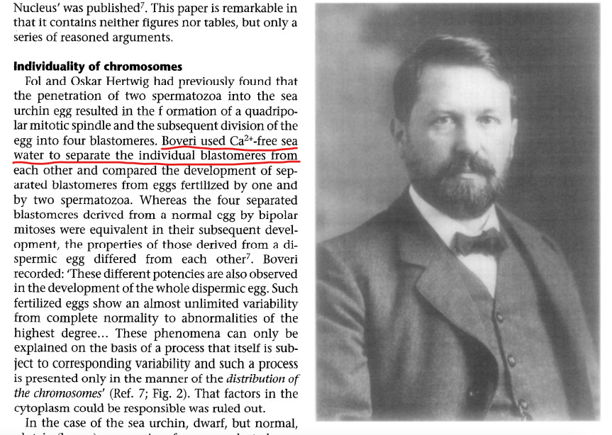
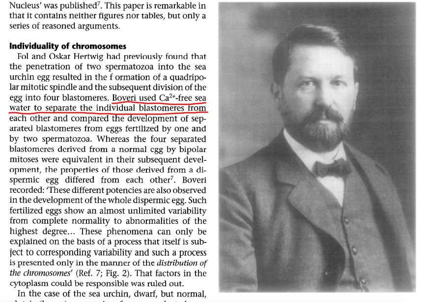
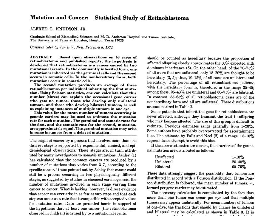
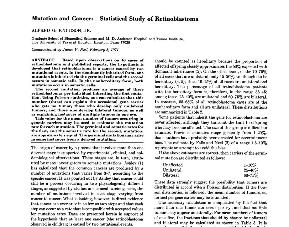
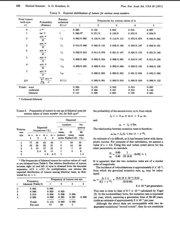
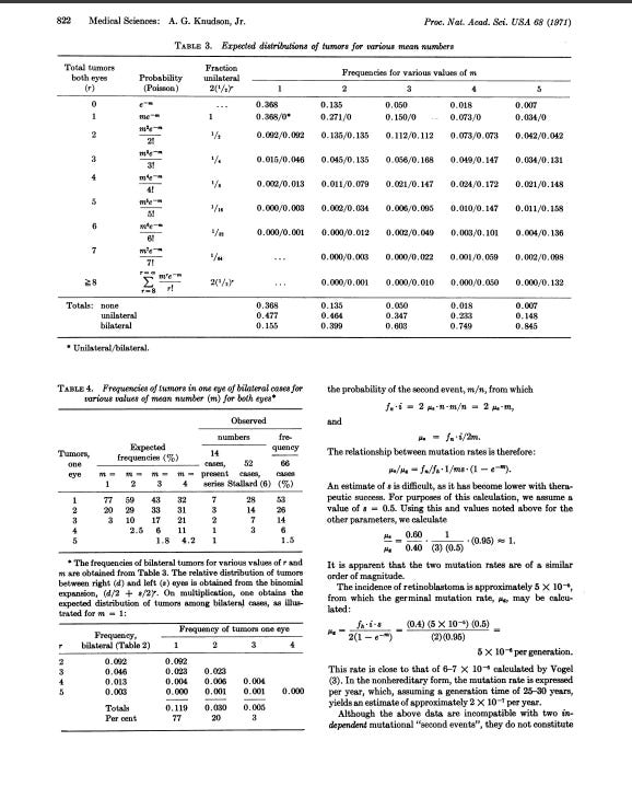
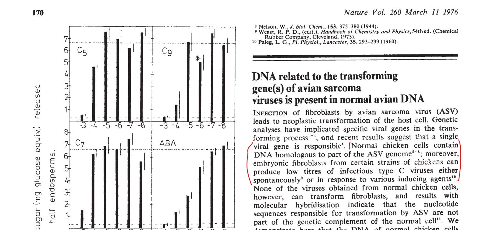
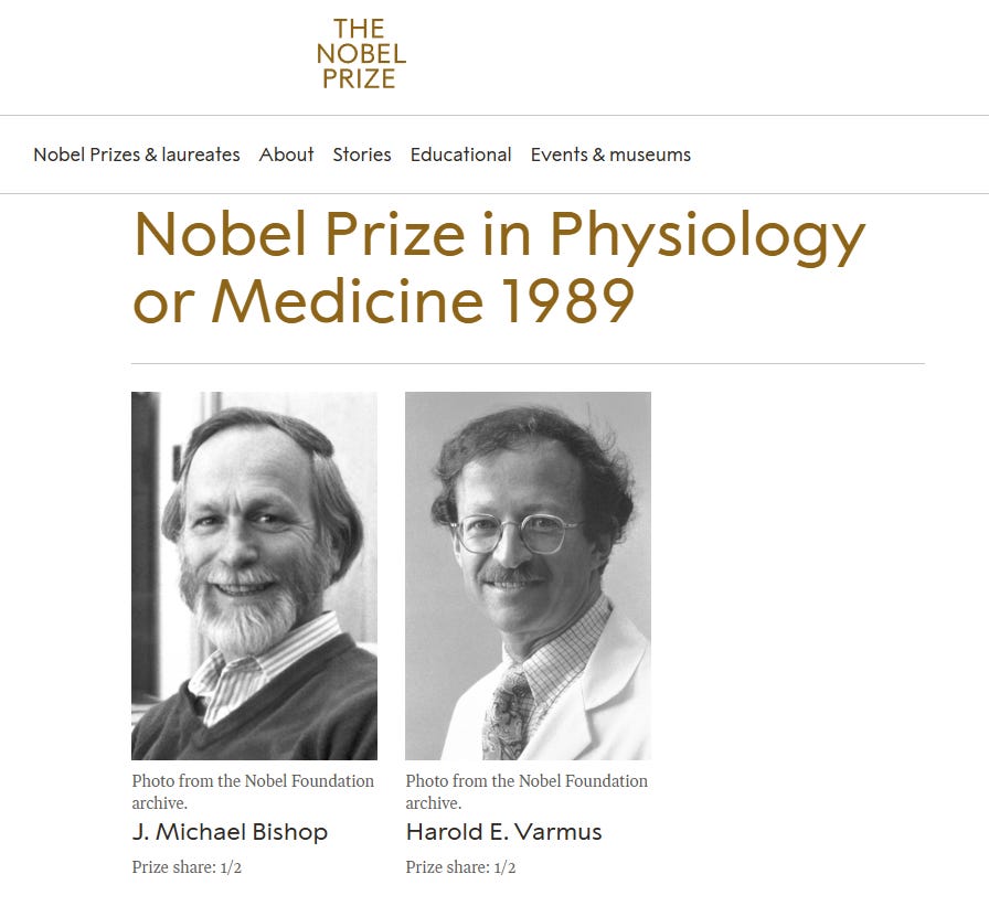
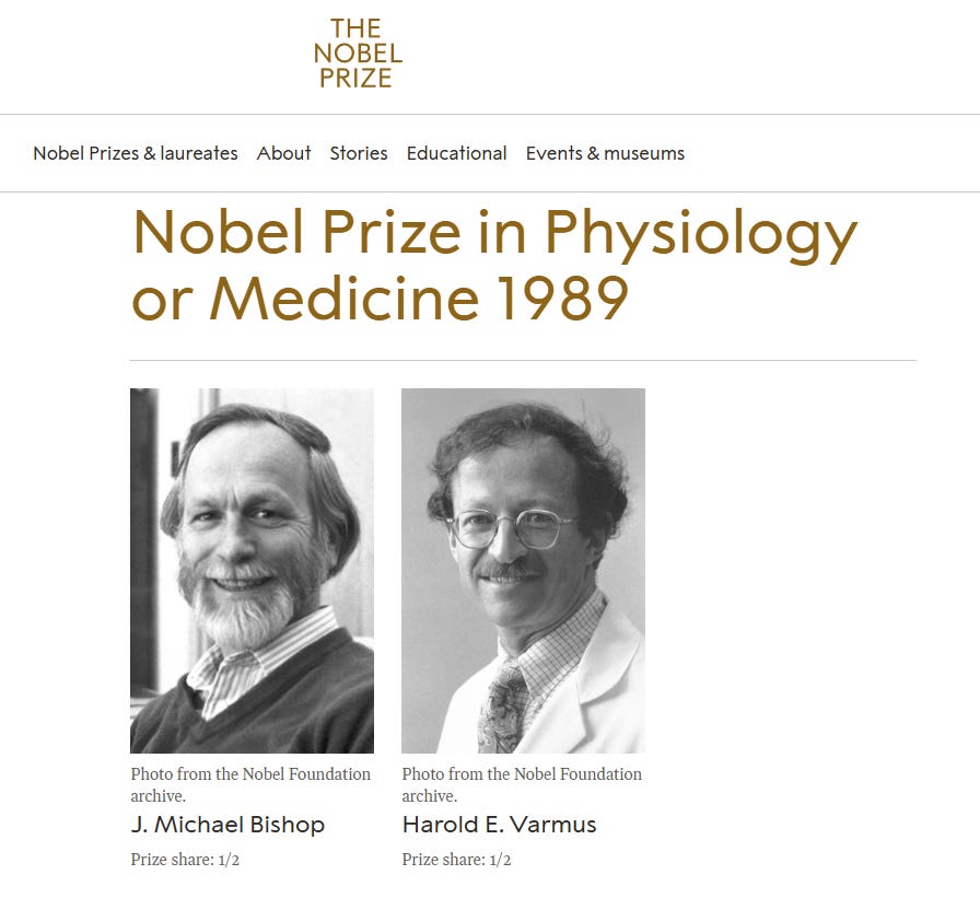
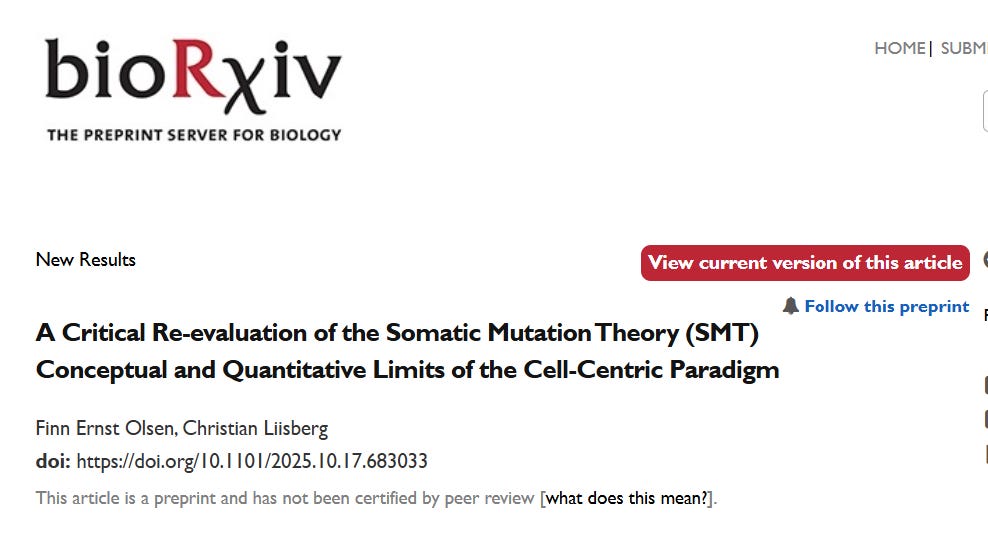
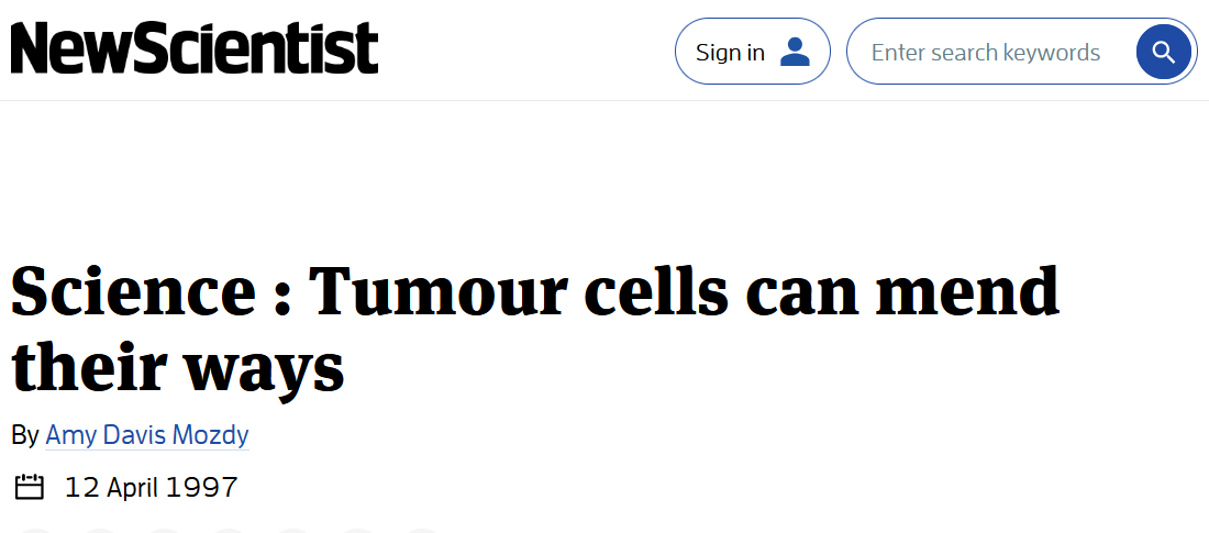
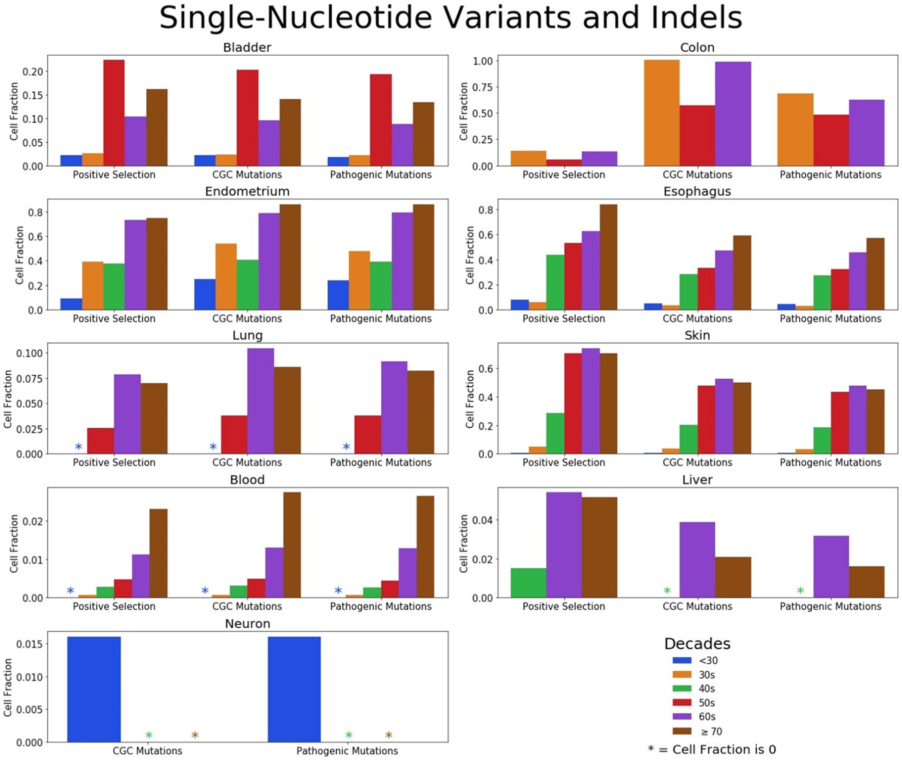
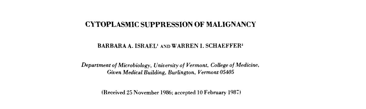
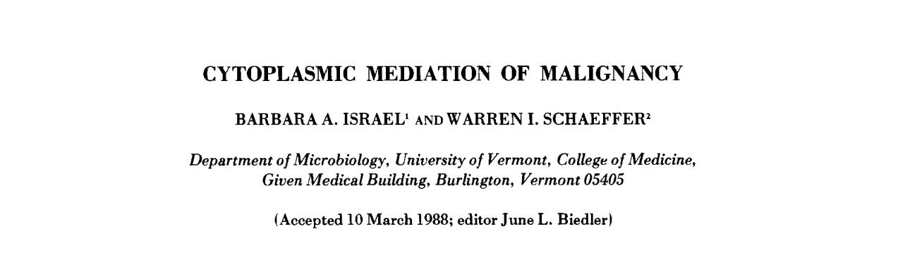
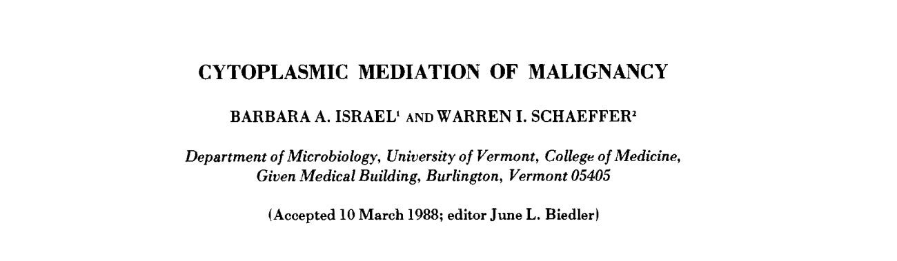
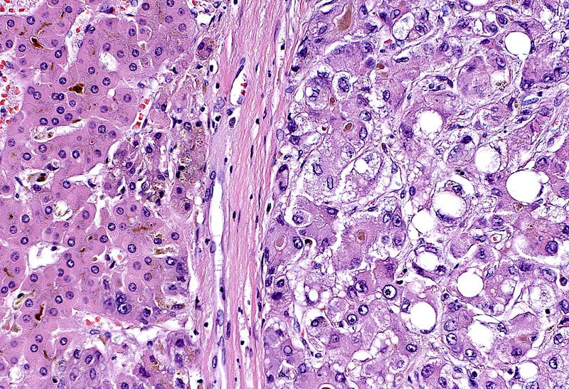
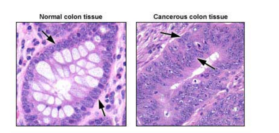
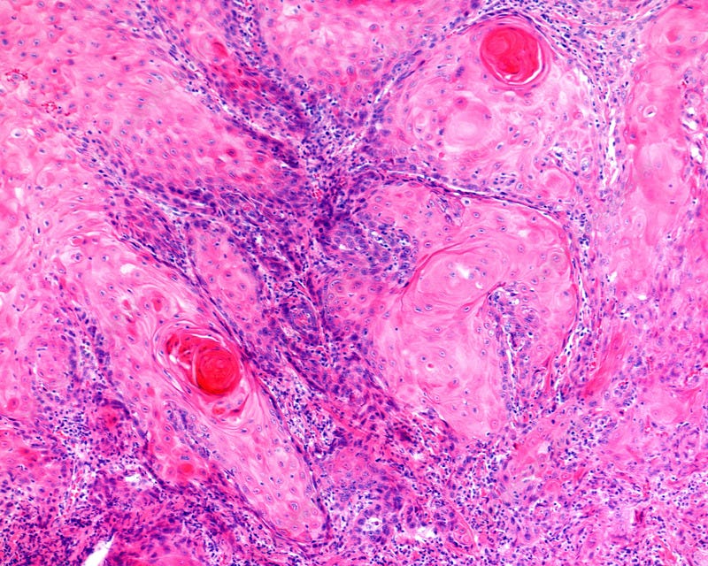
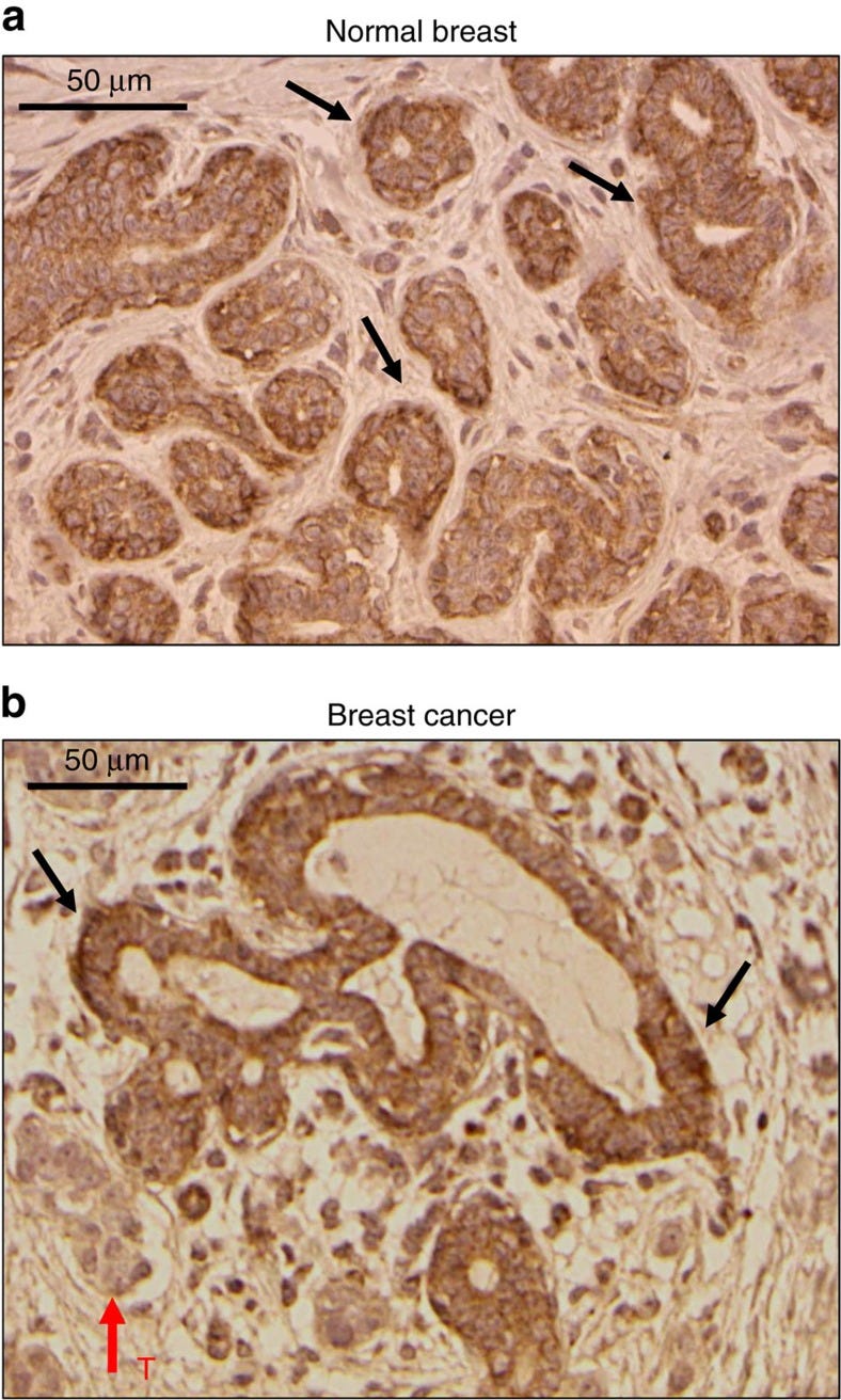
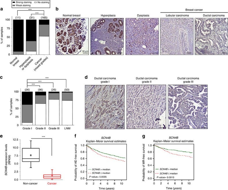
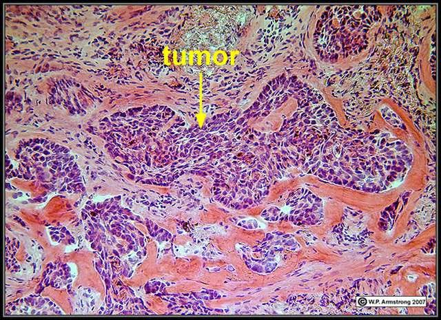
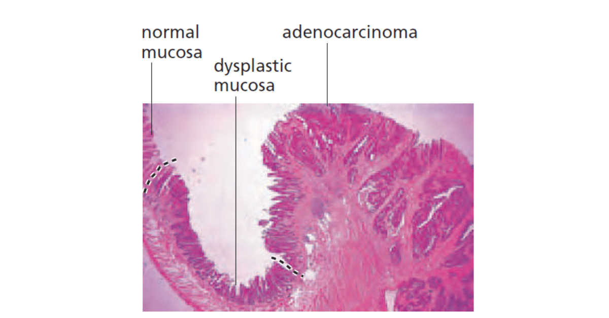
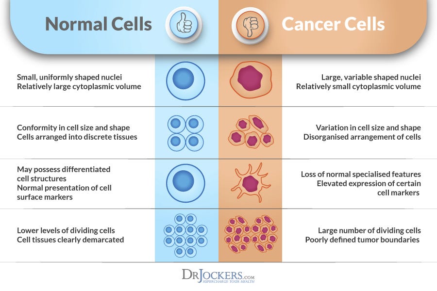
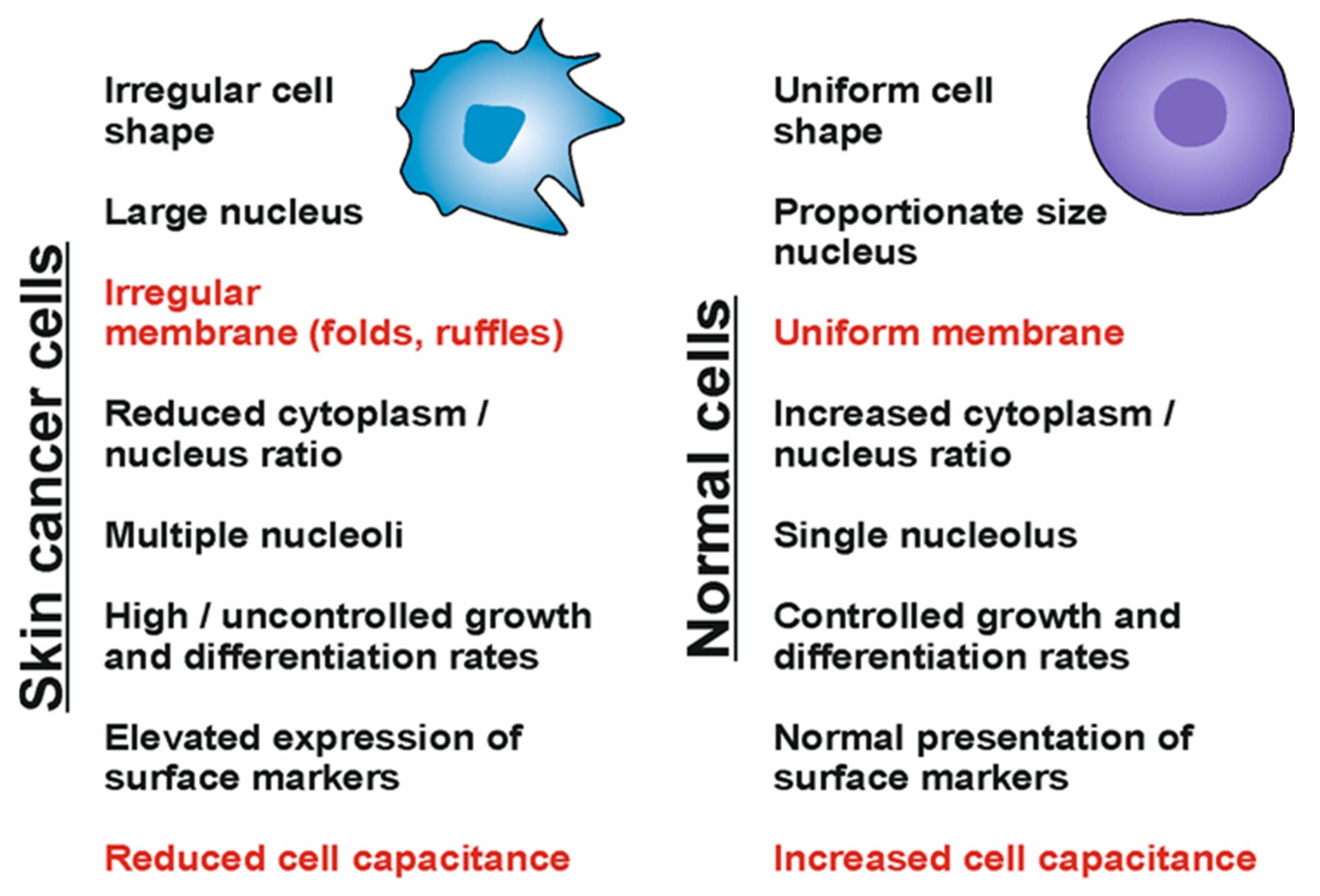
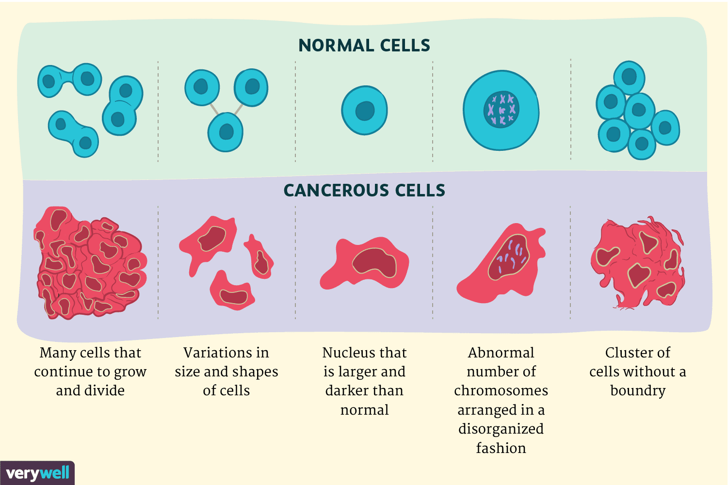
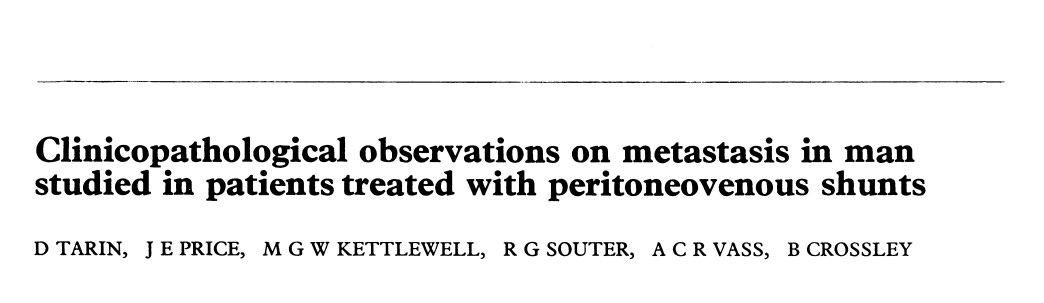
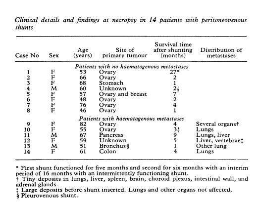
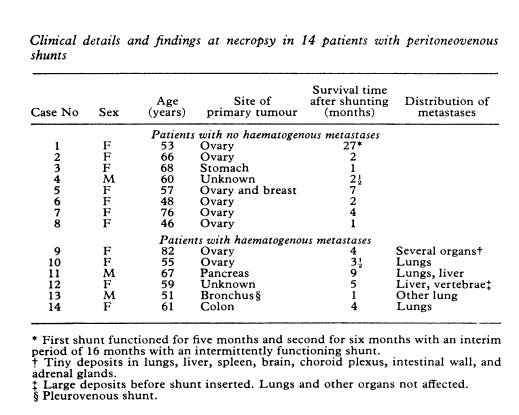
Comments
Nothing yet. Say the first thing.
Sign in to join the conversation.