
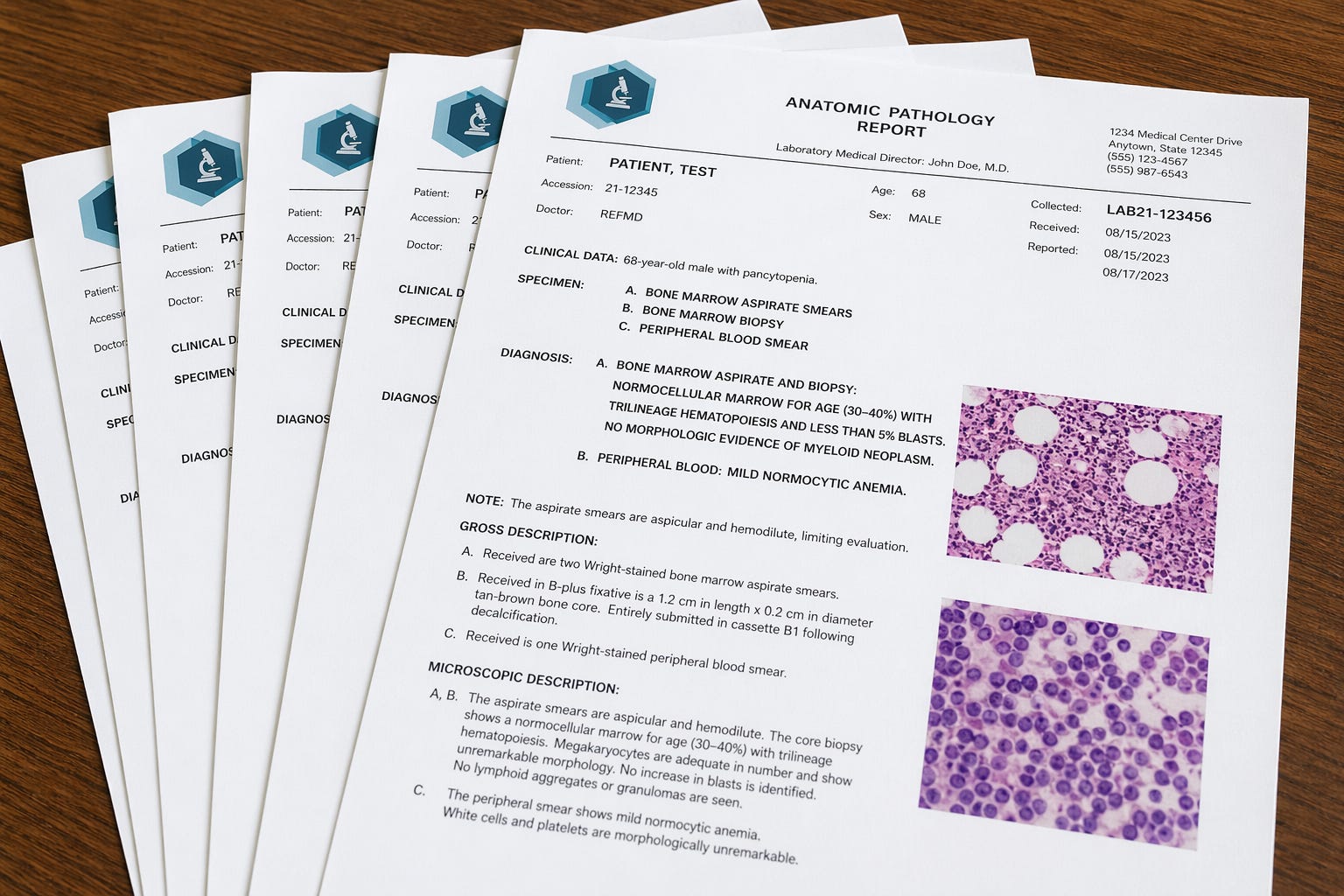
A classic mantra in anatomy and physiology states that “form follows function,” meaning that the structure of cells, tissues, organs, and our entire bodies look the way they do out of necessity. You could analyze medical records in a similar way. If you’ve ever gotten a copy of your pet’s file, you might see notes using the common “SOAP” format:
S - Subjective: Qualitative information, such as temperament, hydration, whether a patient seems depressed or neurologically impaired
O - Objective: Quantitative and physically verifiable information, such as heart rate, temperature, blood pressure, exam findings like a murmur, lab data, etc
A - Assessment: The working diagnosis or opinion based on the “S” and the “A.” This may include comments about the prognosis or expected outcome
P - Plan: Next steps in terms of tests, treatment, procedures, and follow-up
Other parts of the record, such as radiology reports, look completely different. These will usually describe the type of “study” (i.e. X-ray, ultrasound, CT scan, MRI, etc), the images available for review and their quality (in terms of positioning, exposure, and such), and a list of normal and abnormal anatomical structures.
The term “biopsy” was coined by French dermatologist Ernest Besnier in the late 19th century from the Greek roots for “life” and “to see.” It simply means to remove cells or tissue for examination. Biopsies are commonly performed to determine whether a mass is cancerous or not, although there are many other reasons for the procedure. Like the physical exam and radiology reports, a pathologist’s report has its own unique framework, terminology, norms and quirks.
Today, we’ll walk through the parts of a report with examples, and hopefully both vets and pet owners reading this leave with a better understanding of the often mysterious world of biopsy reports!
The term “biopsy” technically applies to evaluation of any cells, fluid, or tissue removed from the body. The specific procedure for large chunks that are fixed in formalin, embedded in wax, and stained with hematoxylin & eosin dyes is called histopathology (sometimes “surgical pathology” is used as a synonym). Direct preps of fine-needle aspirates, smears, swabs, and fluid removed from body cavities is referred to as cytology or cytopathology, and they are usually stained with a modified Wright’s or Papanicolaou stain and viewed at higher magnification (I.e. 50-100x oil lenses).
More recently, you may have heard about so-called “liquid biopsies.” This is a somewhat controversial term to refer to genetic and molecular biomarkers applied to samples from blood, effusions, and urine. The tests are often looking for DNA changes indicative of cancer in circulating tumor cells (CTCs), exosomes, etc. There is a lot less data backing up how useful these tests are, and the reports look at completely different parameters than traditional biopsies, so I won’t be addressing them here.
The overall format of a pathology report—with a few minor exceptions—has remained largely unchanged for over 100 years. The basic parts consist of a descriptive section (think of this as a combination of subjective/objective from SOAP), a diagnosis or interpretation (the “A”), and comments (the “P”). These reports also contain patient ID information and some details on the history the physician or vet sent in with the sample. This is a mock-up from the College of American Pathologists:
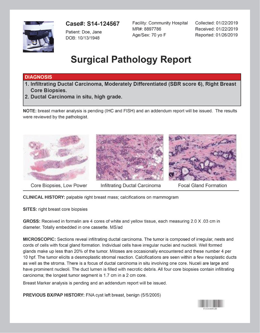
The description is the meat and potatoes of a biopsy report, where the pathologist documents everything they see without providing an opinion. You can think of it as the “just the facts, ma’am” section. One thing that can make the description section intimidating for general practitioners, patients, or pet owners to read is that it’s often filled with a lot of specialized jargon. Looking up unfamiliar terms with Google or ChatGPT can help decipher what’s going on. It’s also nice when pathologists explain less common terms using parentheses or other formatting (I often do this).
The microscopic description is common to histopathology and cytology, and documents what types of cells/tissues are present on the slide at high magnification, if they look normal or abnormal, presence of bacteria or fungi, foreign material, etc. There are often a lot of references to size, shape, color, and specific appearance. On cancer reports, the description usually mentions “margins,” or how much space there is between normal tissue and tumor (to see how complete removal was). The length of this section can range from a sentence or two to multiple paragraphs, depending on the type and complexity of the sample. For histopathology reports, there is also a gross description in addition to the microscopic. These comments describe how the tissue looks with the naked eye before processing, and might be something like: “2x3 cm pale, tan, rubbery, subcutaneous mass.”
In the beginning, a written description was necessary to provide a legal record of the sample because pathologists often work in remote facilities away from their referring physicians or veterinarians, attaching photos was not technologically feasible, and the original slides may not have been stored longterm. Today, the combination of regulations mandating minimum sample retention times (often years) and ease of adding images to reports might seem like it makes this redundant. However, in my opinion (and that of other pathologists), it still adds value. For one thing, it can be used for consulting or second opinion requests. For another, it also contains valuable information in the medical record for data mining in research and clinical trials1.
This is where the pathologist puts a label on the disease process and decides if the sample is most consistent with cancer, inflammation, an infection, or something else. You can think of this as basically the same as a diagnosis, although some veterinary labs call it an “interpretation” and suggest the ultimate diagnosis is up to the attending clinician who is actually seeing the patient. It’s a pedantic difference in wording to reduce liability risks, and one that I’m not sure provides much protection, but it is a common industry practice.
In some cases, the interpretation may be short and sweet, like “canine cutaneous histiocytoma” (a common benign skin tumor). Other times, it might be more of a “morphologic” diagnosis, or a string of anatomical and pathological terms along the lines of: “Moderate lymphoplasmacytic to mildly neutrophilic duodenitis with multifocal lacteal dilation and mucosal erosions.” Putting that into plain English: the location is the duodenum (small intestine), the suffix“-itis” means there is inflammation, and the infiltrate is composed of neutrophils, lymphocytes, and plasma cells, along with some damage to the surface lining and swelling of internal structures → it’s basically Inflammatory Bowel Disease.
You might ask, “Why doesn’t the pathologist just say IBD and be done with it??” The reason is that each one of those modifier words characterizes the specific “flavor” of the patient’s IBD, and might carry different treatment or prognosis implications.
For some really simple lesions, there may be no additional comments. However, it is quite common for pathologists to use this field to do one or more of the following:
Ask for more clinical details or follow-up
Discuss any quality issues (limitations based on small sample, crushed tissues, etc)
Recommend additional tests, especially if the diagnosis is in doubt
Provide information on prognosis, explain tumor grading
Reference studies to back up their thought process (especially if it’s a rare disease)
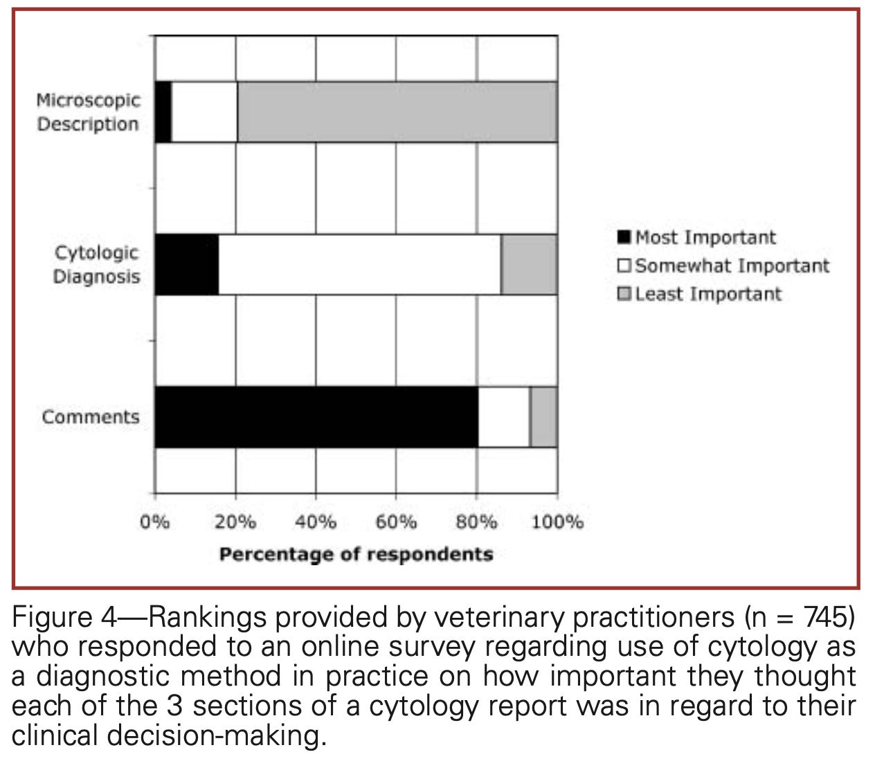
The comments section is often rated as the most important part of the report by clinicians, as in this study:
A common situation where I use the comments is to explain my grading decisions for canine mast cell tumors (MCT), because the system is relatively new and comes with a lot of caveats and exceptions:
Another one is with lymph node samples suspected to be lymphoma. If the slides are not a slam dunk for cancer, I may recommend confirming with a molecular test like PARR. Even if I am sure of the diagnosis, following up with something like flow cytometry can give an oncologist more information about how to treat it.
Finally, as I put it in my semi-satirical post The Laws of Pathology: “The length of the comments is inversely proportional to the confidence of the pathologist”. If I am writing a novel-length treatise, you should know that this is something really bizarre!
There are a variety of additional sections that might be added to your report. Digital photos of the lesion have become relatively common in veterinary biopsy reports. A picture is worth a thousand words, and even a few can say more to a GP or patient than the long description ever could.
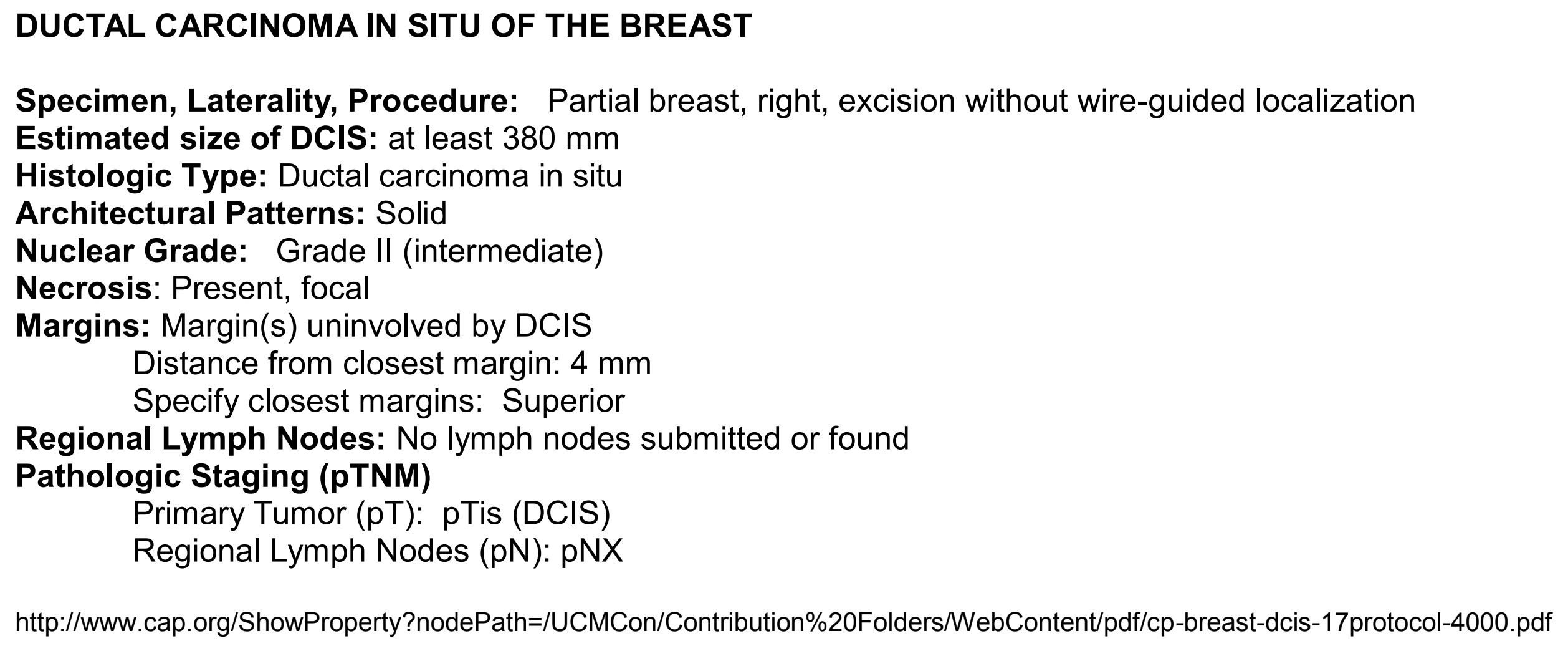
Cancer cases often include a “synoptic report,” or a checklist of relevant features for that specific tumor. An example might look like this:
Then, there are two sections that sound almost identical, but are quite different:
Addendum: This is when new information is added. A common situation is when “special stains” are requested. The pathologist might initially look at a lymph node and be unsure if it is reactive or cancer, so they order immunohistochemistry (IHC). Then when it comes back, they will write an addendum, such as “All of the lymphocytes were B cells, the diagnosis is B-cell lymphoma.”
Amendment: An amended report is when something is changed from the original. This can be to correct a typo, but may also indicate a more substantial error that was caught and remedied. Many labs track pathologist amendment rates as a quality benchmark.
The surgeon Richard Selzer once wrote “Autopsies give us the facts, but not the truth.” The same could be said for biopsies. Pathologists are taking an extremely small piece of a patient’s body at a single point in time and trying to figure out what’s wrong with them based on that. They can be led astray for numerous reasons, ranging from too little tissue sampled, to damage to the specimen, to a lack of relevant medical history, and more. Some diseases are tricky to diagnose with a biopsy and may require imaging or other types of tests. Finally, the subjective experience of illness may not always match what is seen under the microscope: One patient may have minimal inflammation on their biopsy slide, but be in serious pain, while another could have a sample that looks markedly abnormal but experience no symptoms.
The upshot is reports may be written defensively, with a lot of hedging language around probabilities and uncertainty. This is likely much more common in large diagnostic labs like Quest or Labcorp on the human side, and Antech/IDEXX/Zoetis on the veterinary side. Sometimes having the primary care provider contact the pathologist can clear up any confusion and lead to a better outcome.
To handle the inherent ambiguity of pathology, it is often necessary to rely on multiple different tests before taking any serious action like chemotherapy, surgery, radiation, etc. Many diagnostic labs already have formal and/or informal consulting processes in place for serious diagnoses, but if you have any doubts or concerns, you or your physician/vet can request a second opinion.
What has your experience been with biopsies for your pets or your own health? Leave any comments or questions below!
—Eric

“How to Read Your Pathology Report” — College of American Pathologists
“Understanding Your Pathology Report” — American Cancer Society
Author’s Note: The commentary above represents my personal opinion and does not necessarily reflect the viewpoints of any current or previous employers.
It would not be practical to manually review all of the biopsy slides in a large clinical trial, so researchers rely on the pathology reports to properly classify patients
Comments
Nothing yet. Say the first thing.
Sign in to join the conversation.